
Last modified on 09 July 2013.
Introduction:
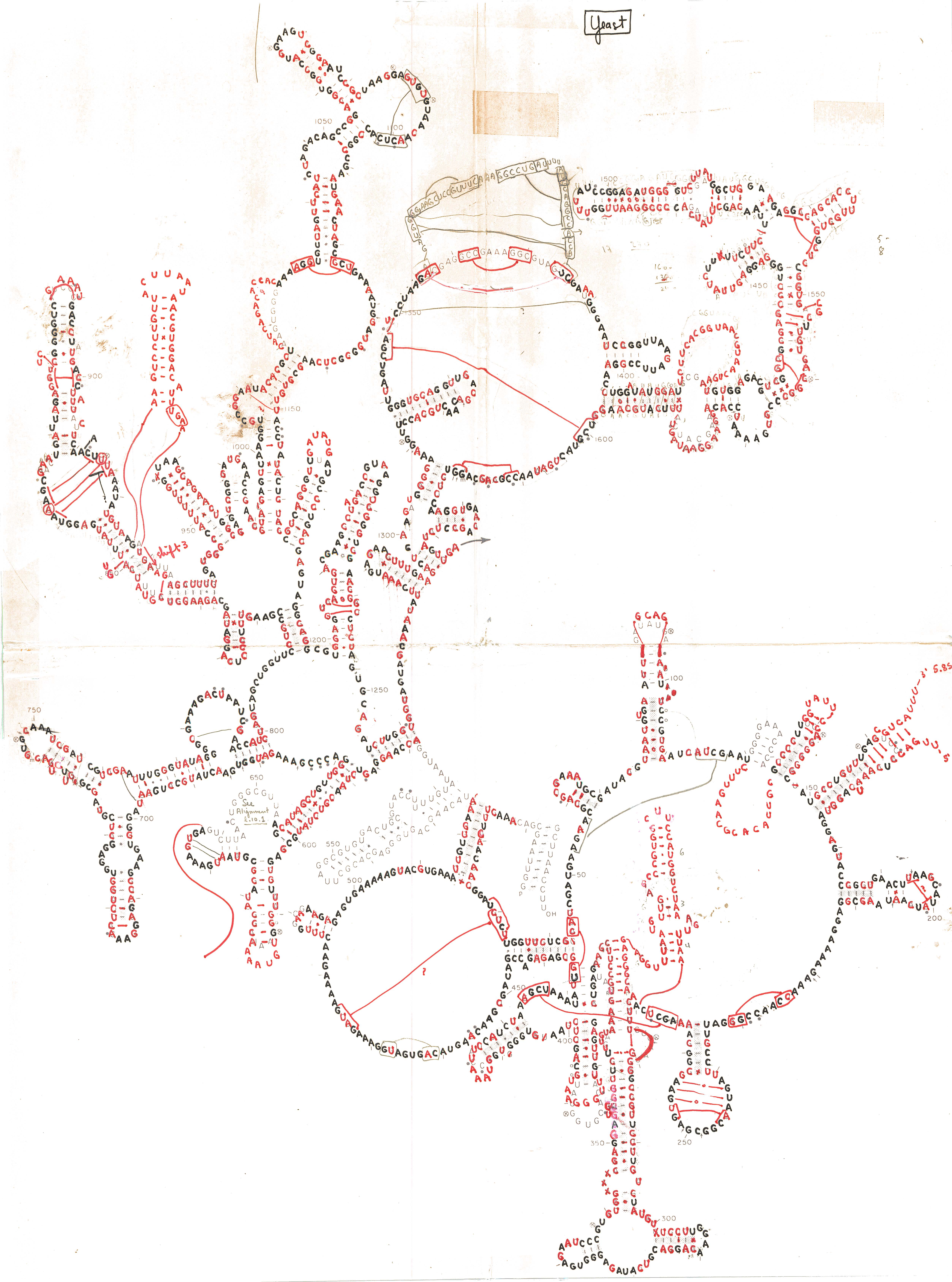
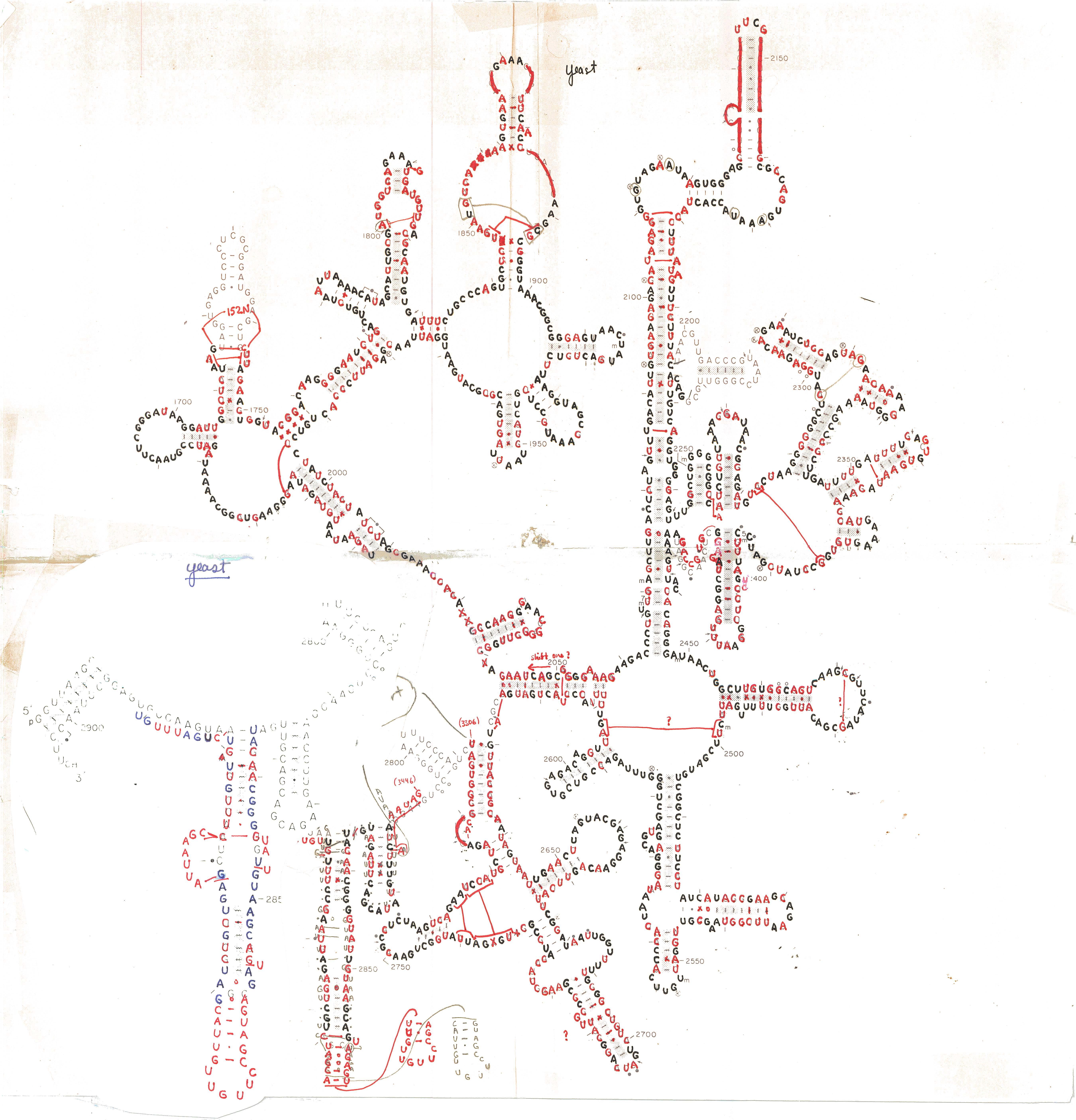
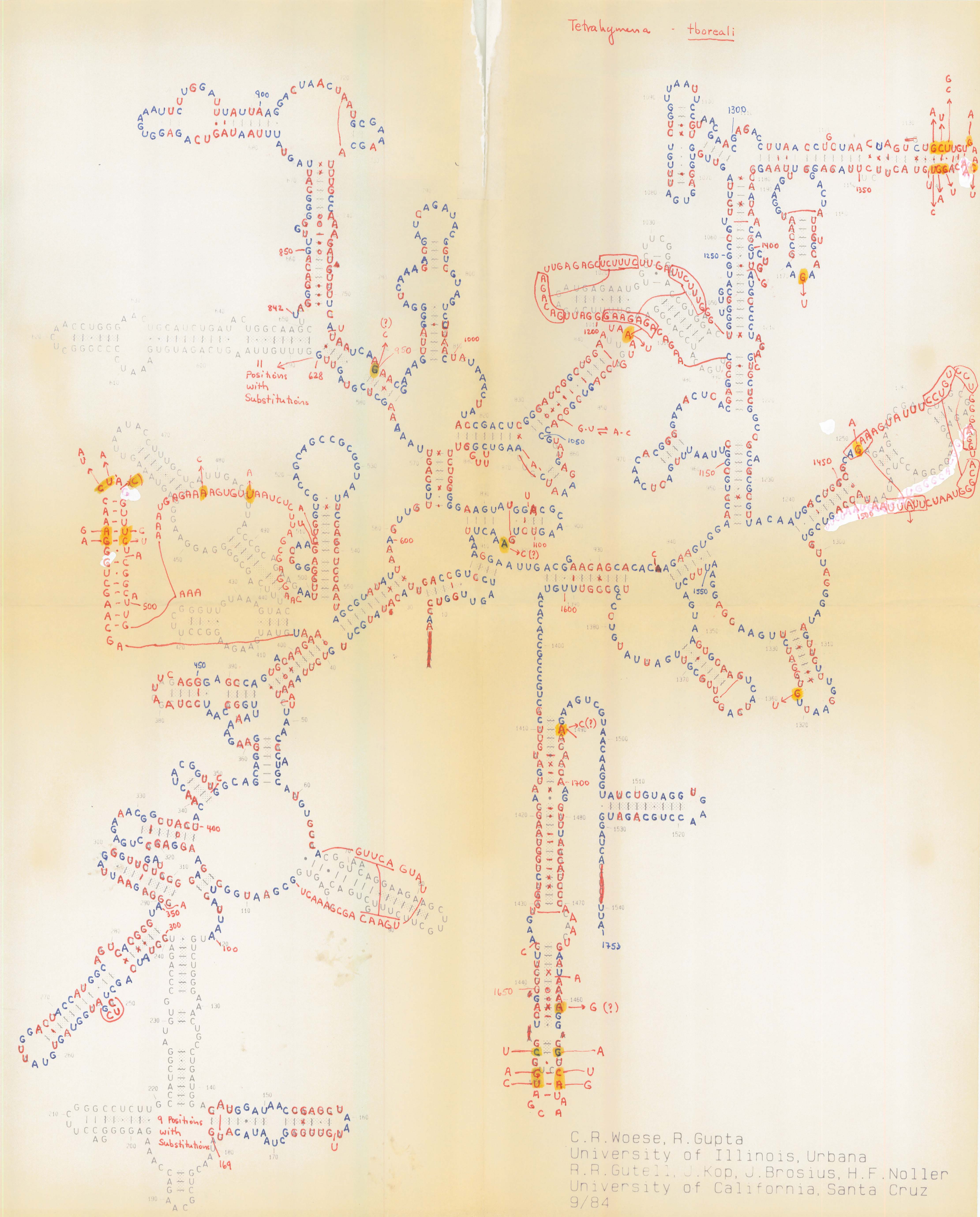
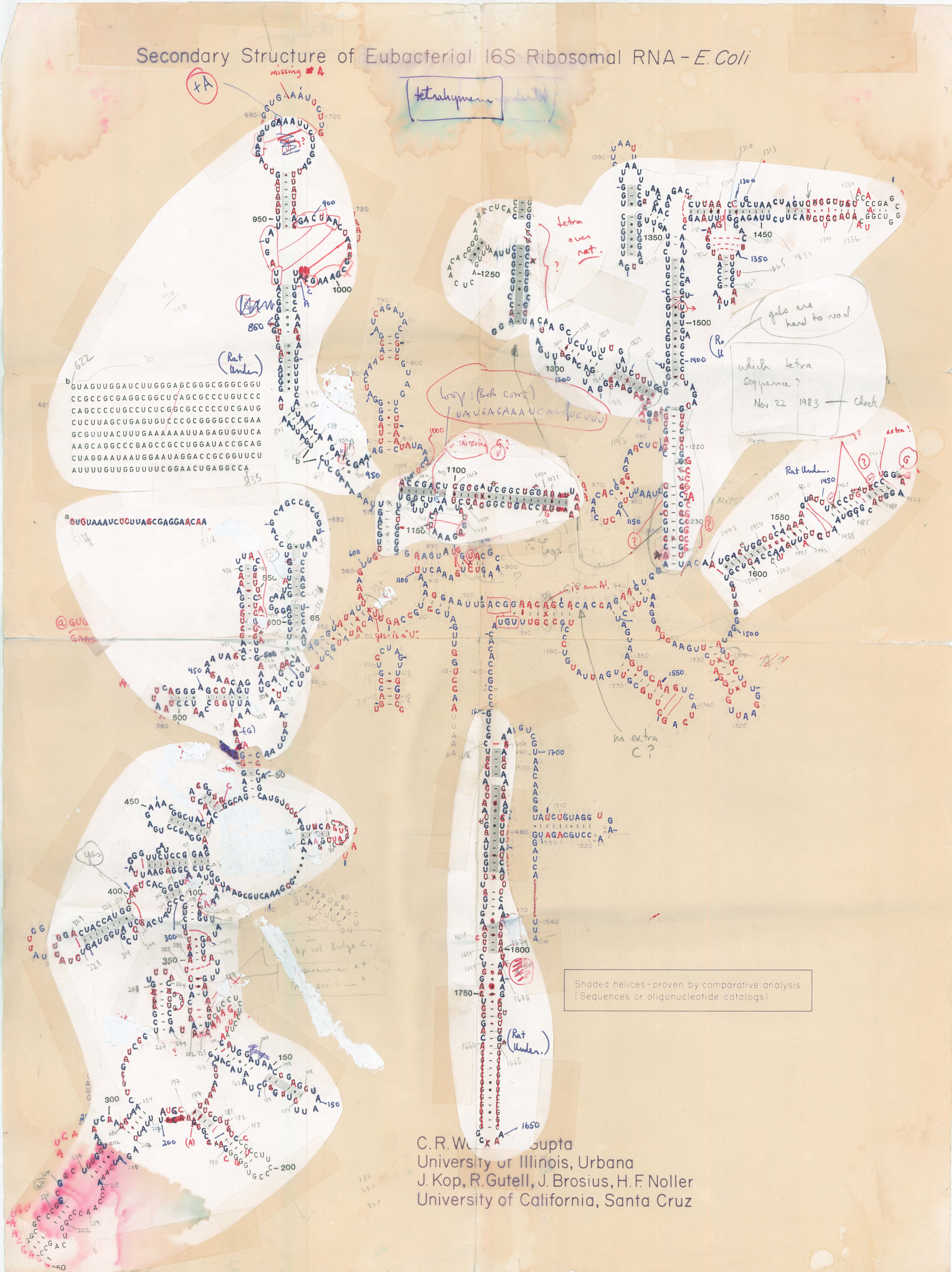
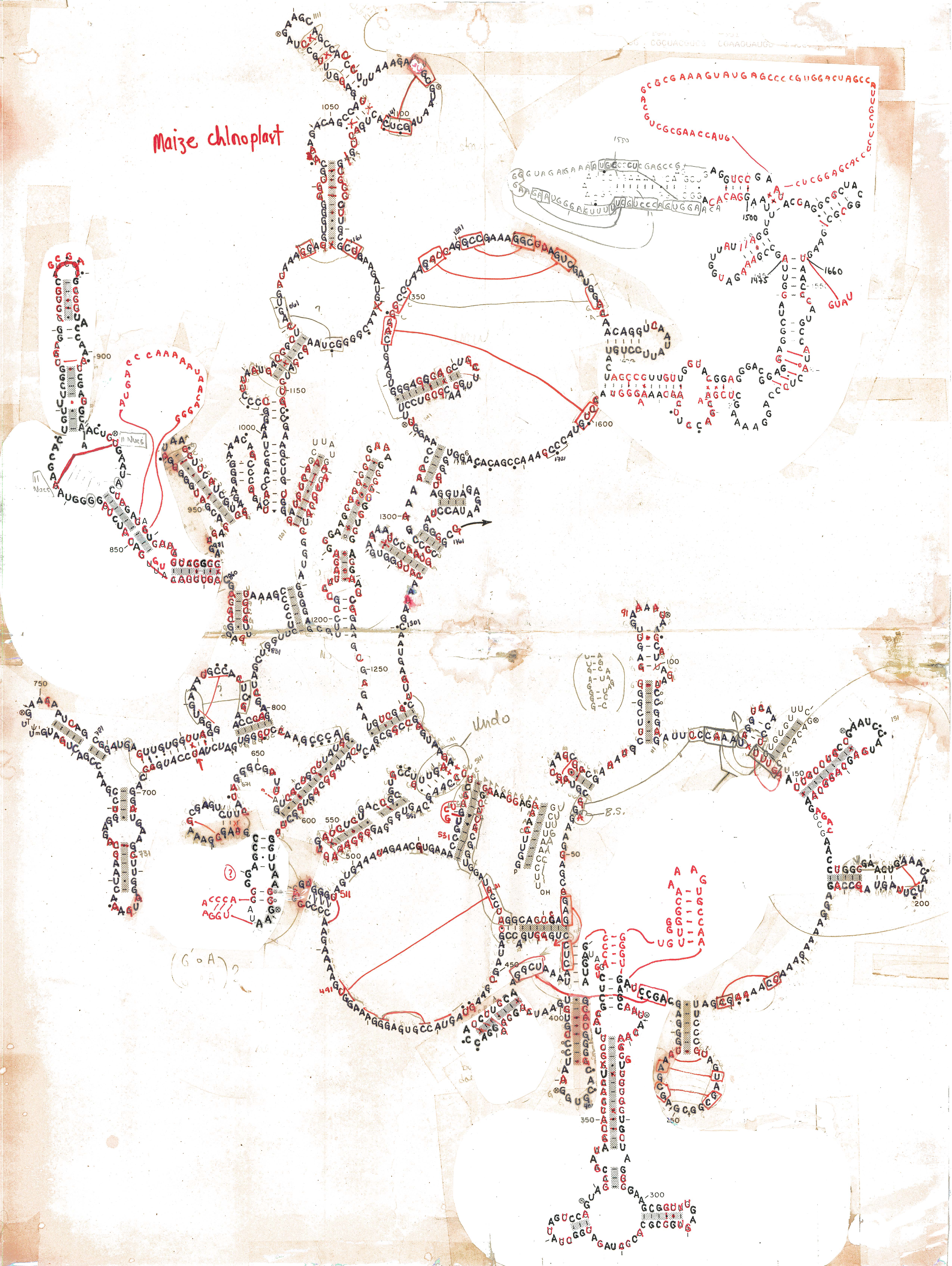
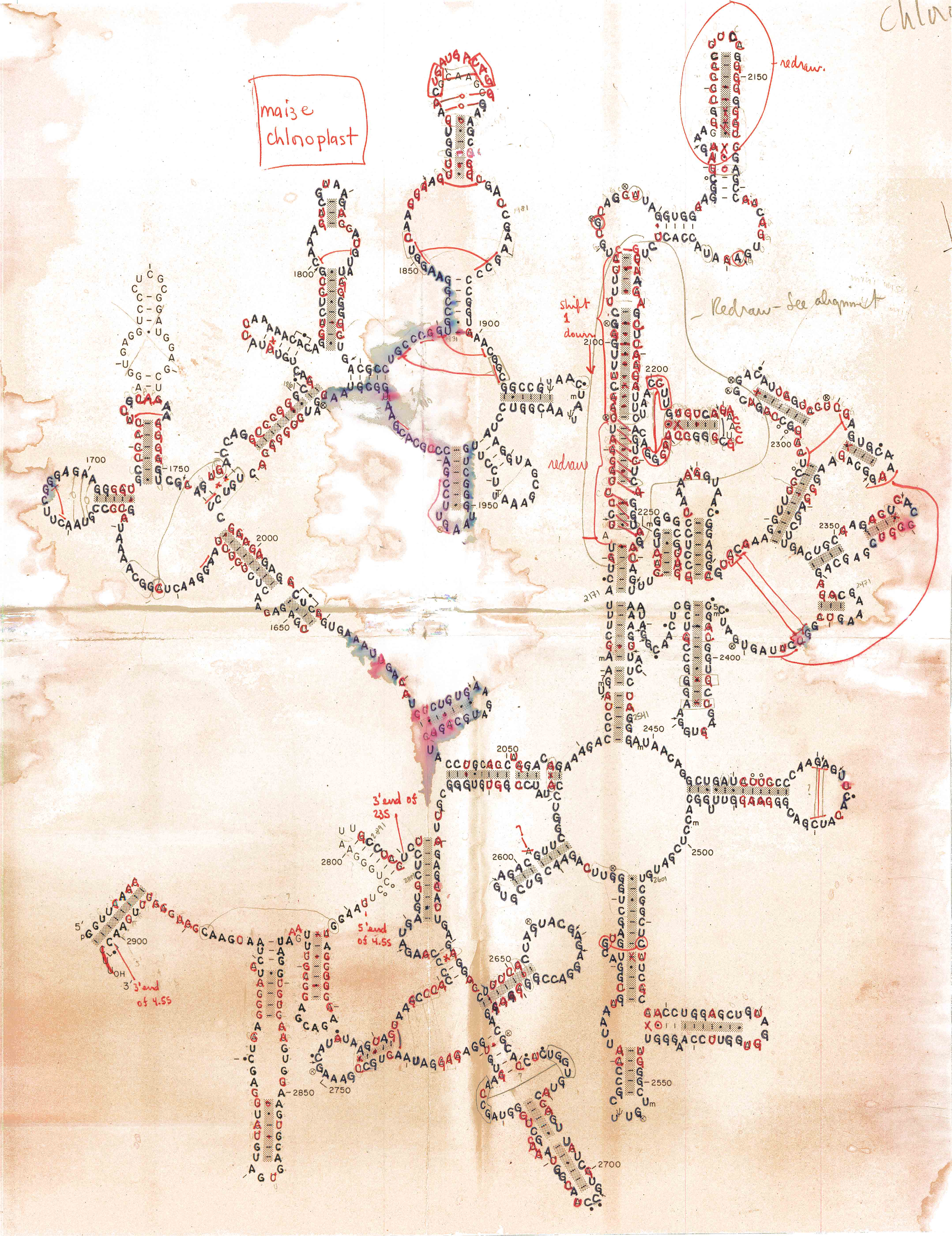
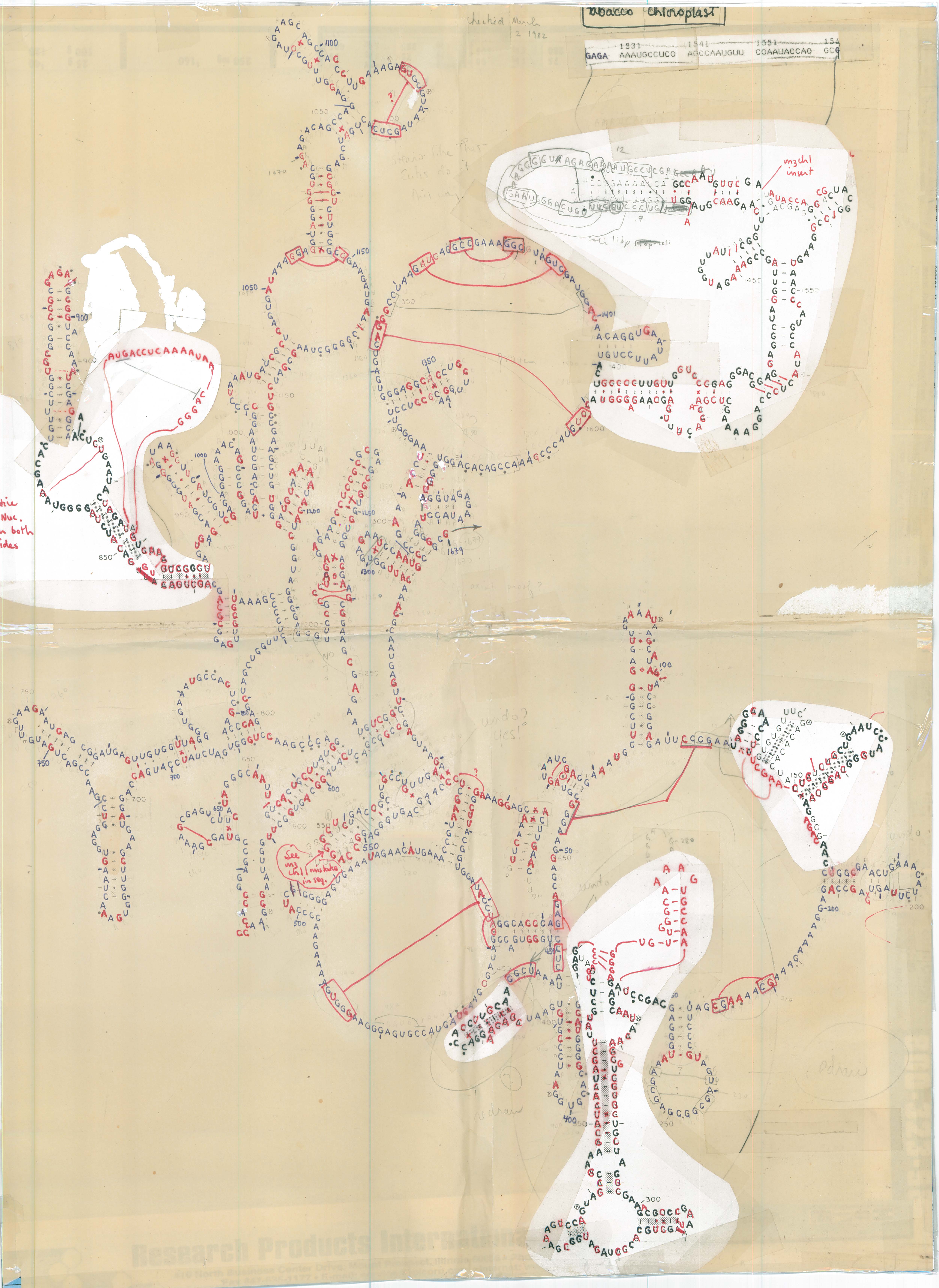
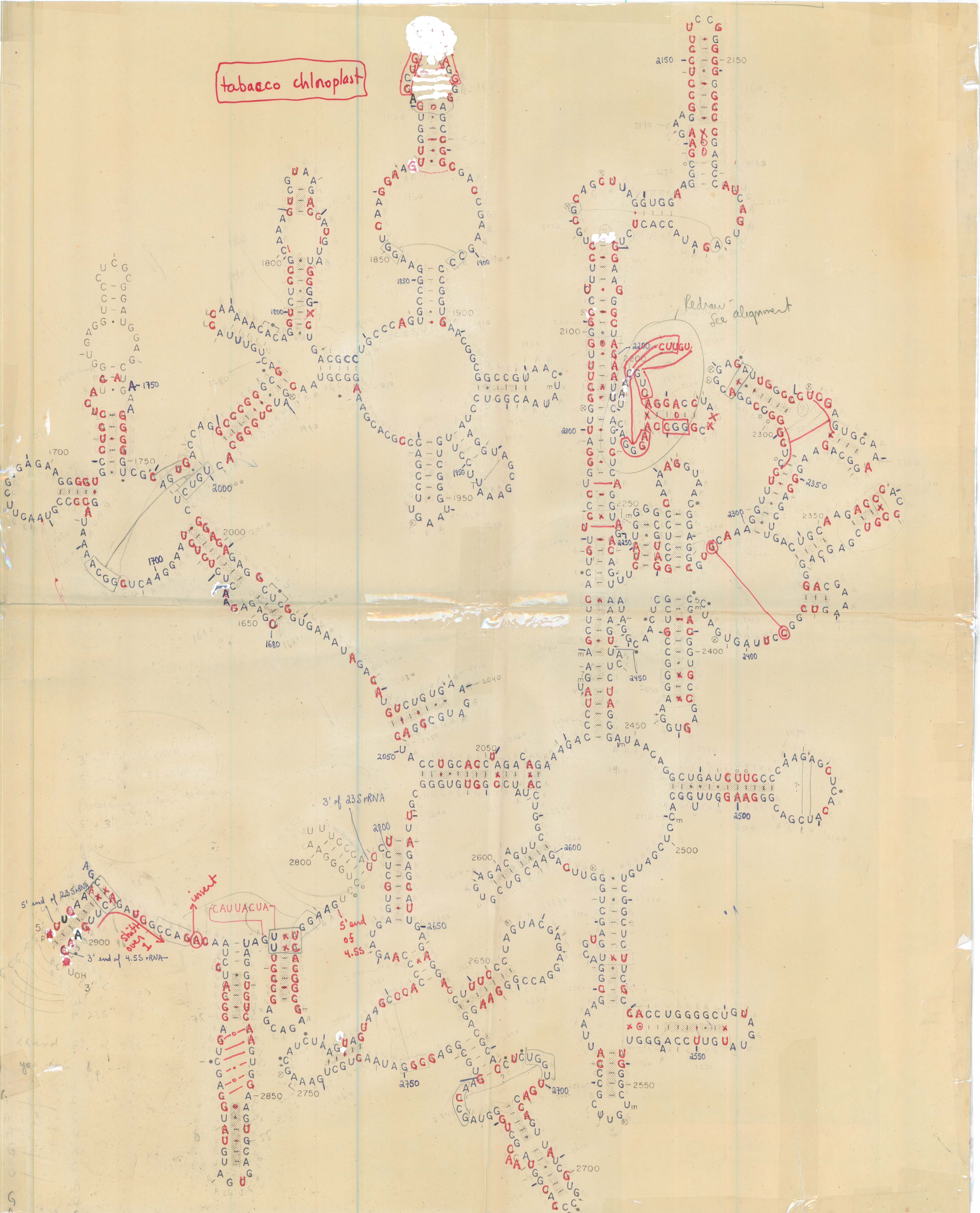
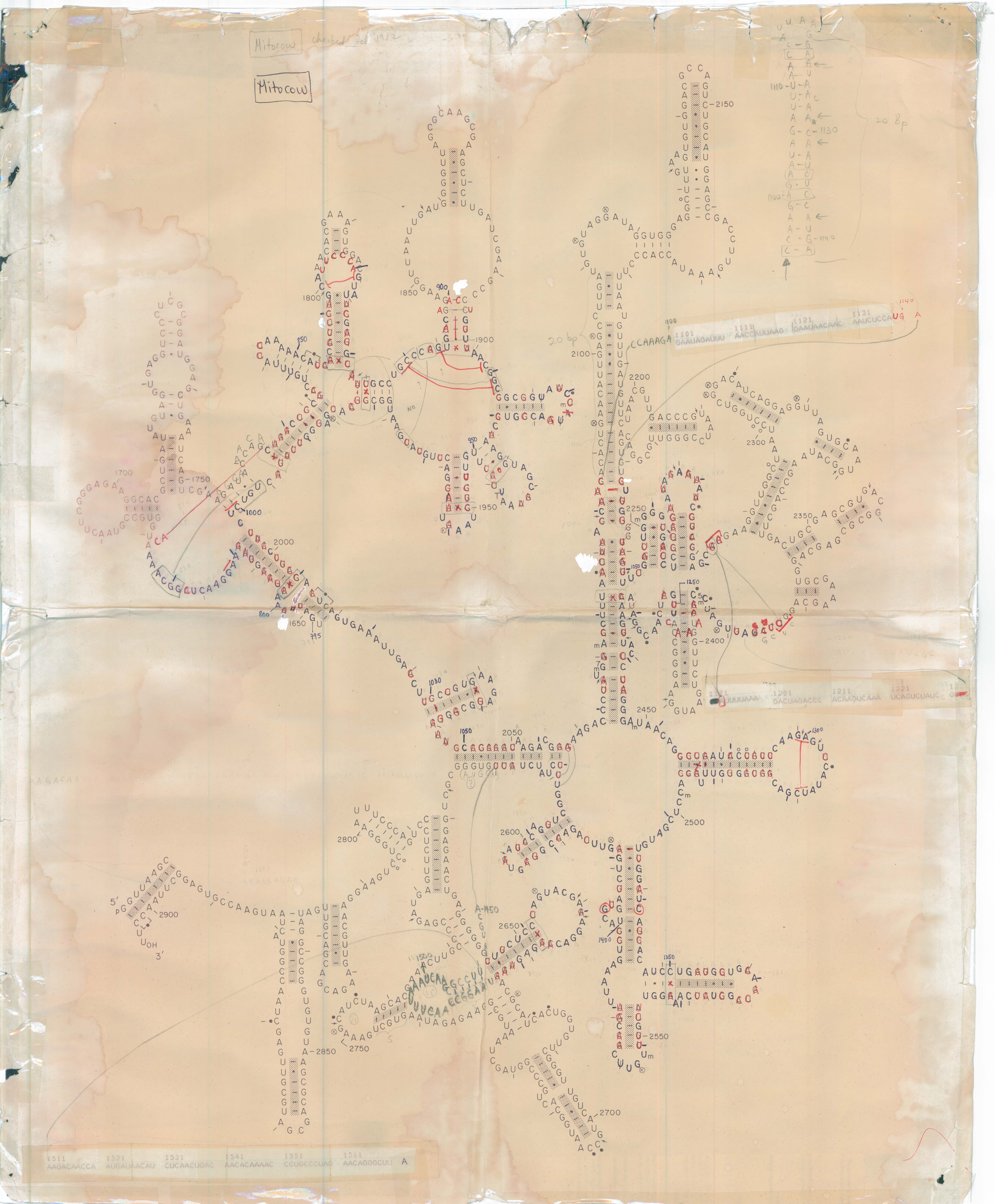
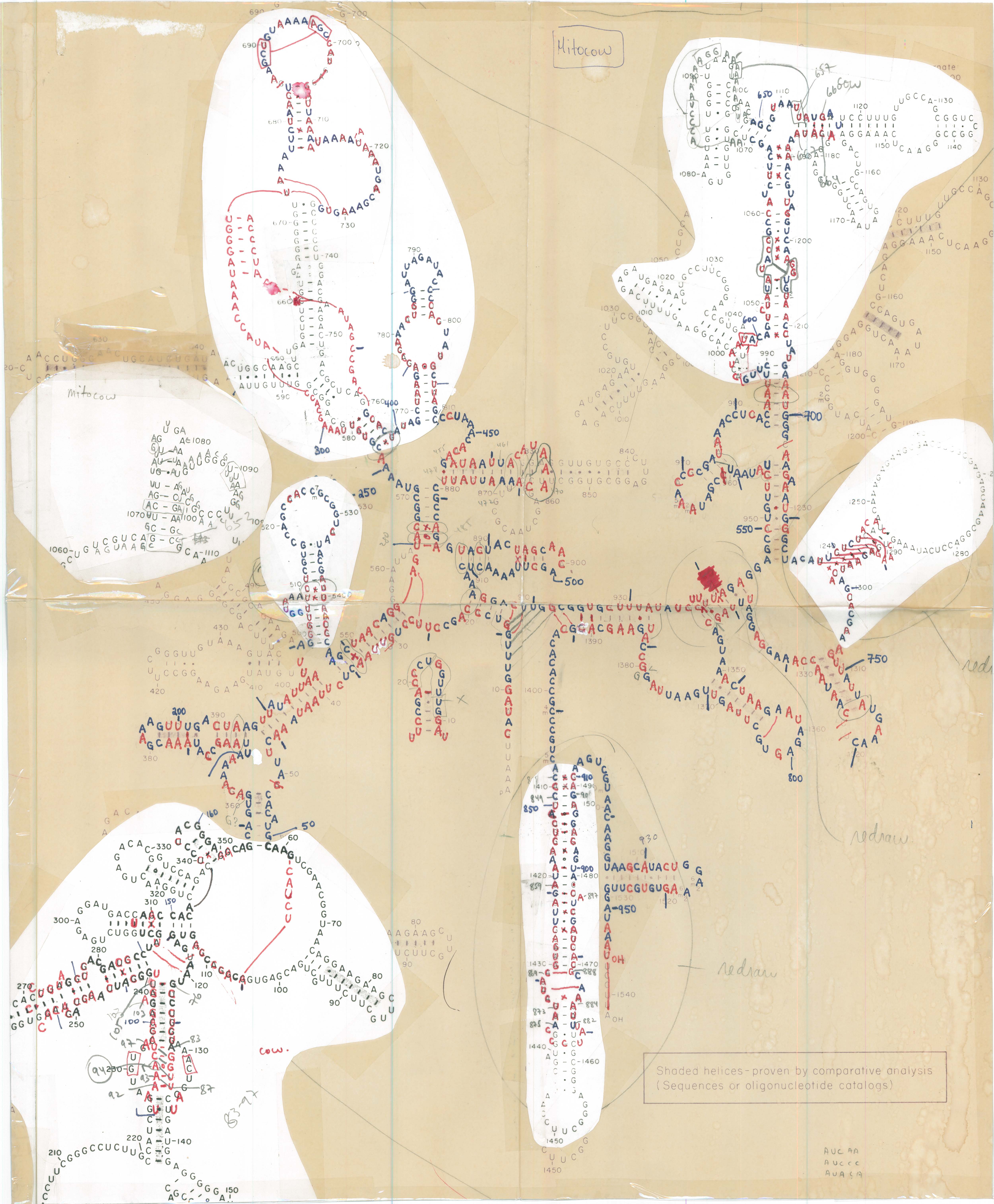
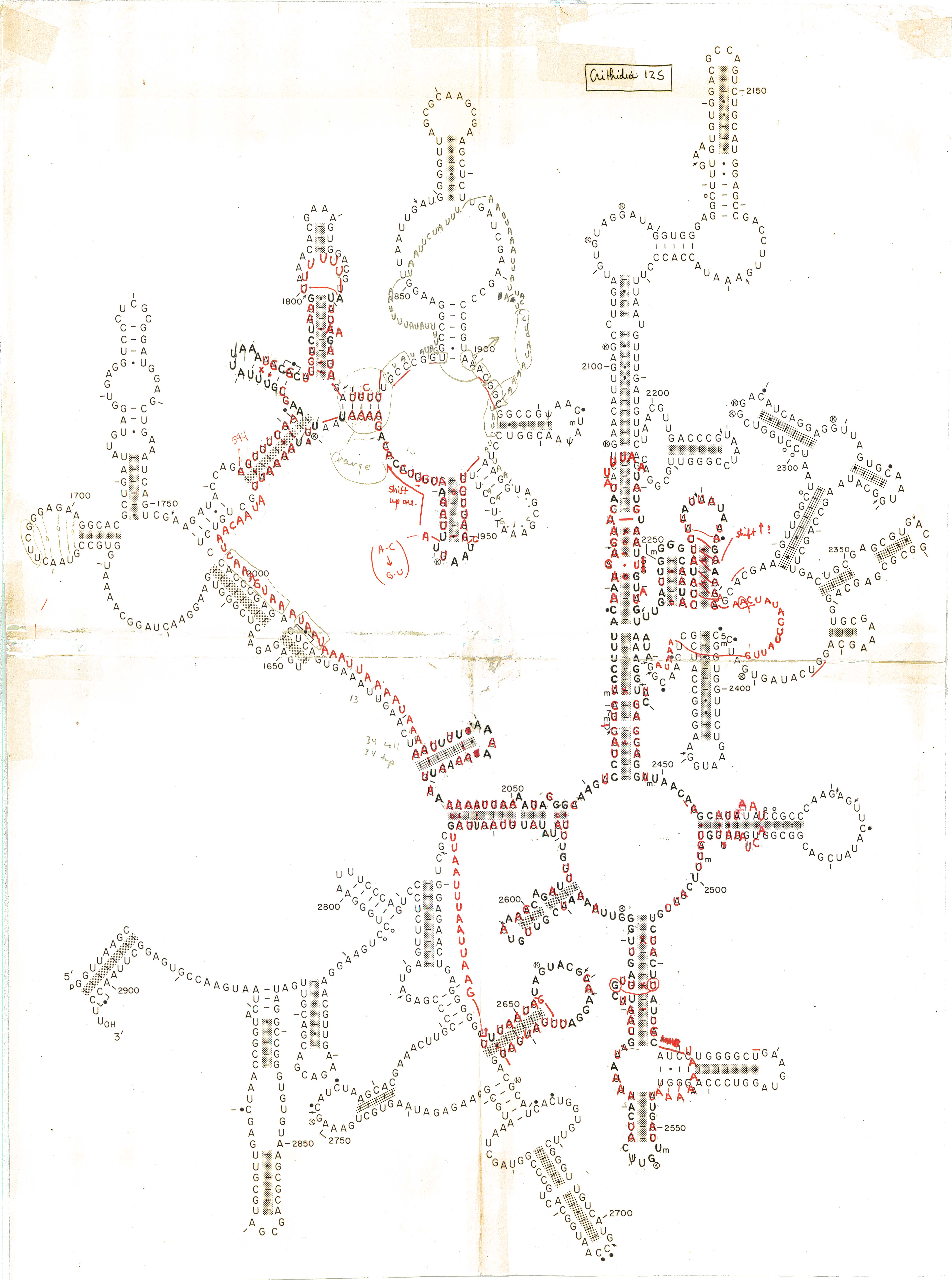
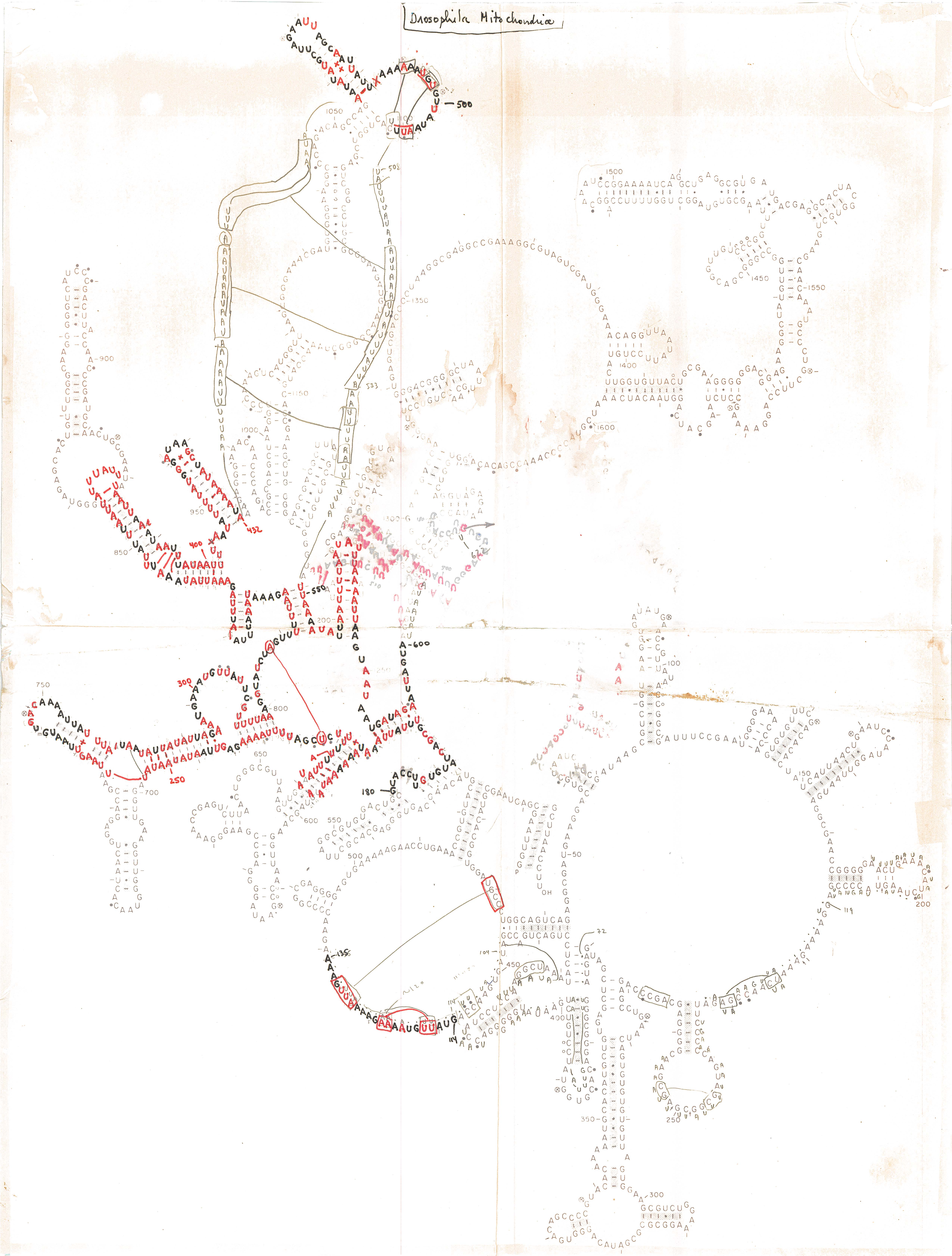
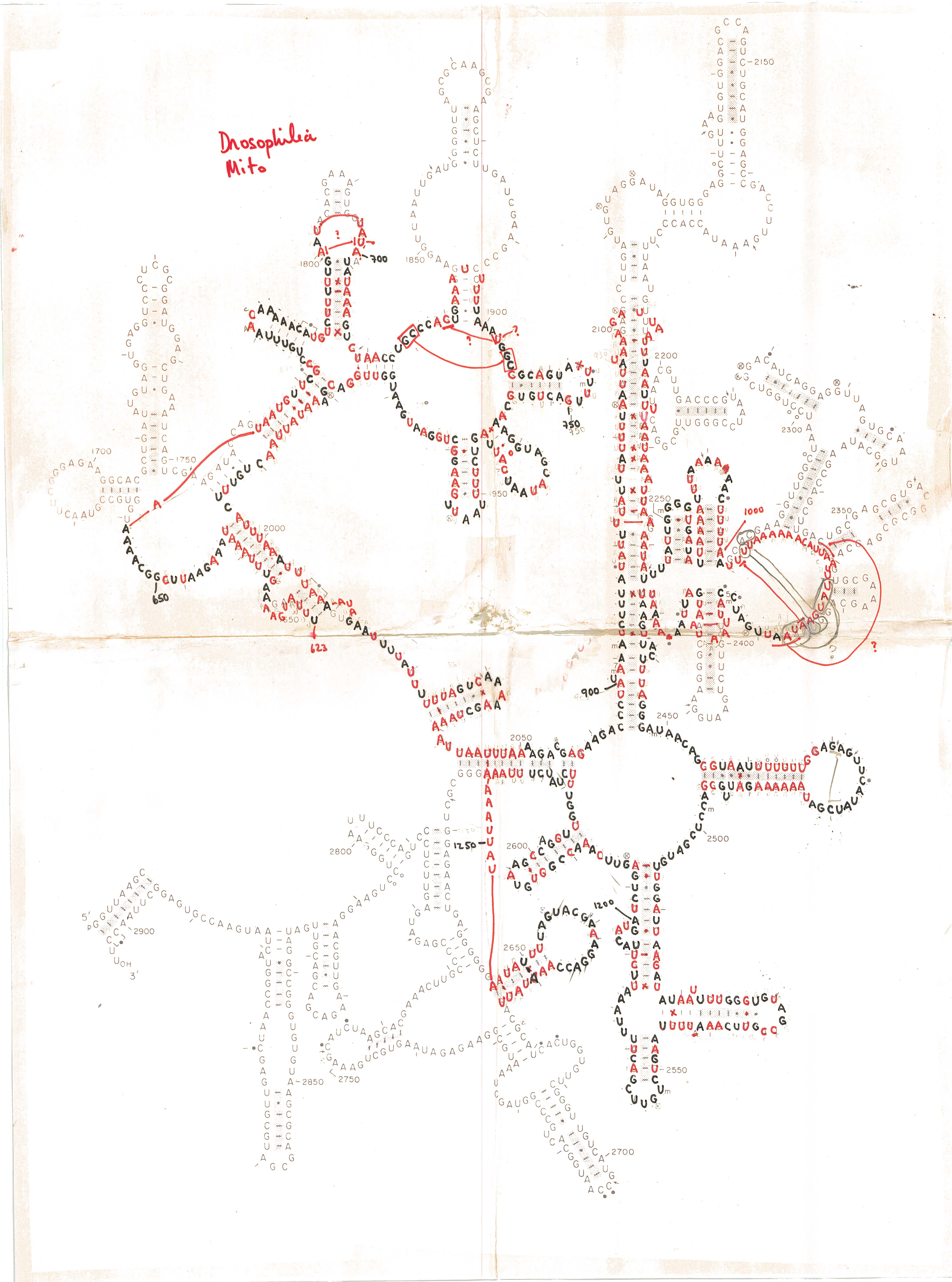
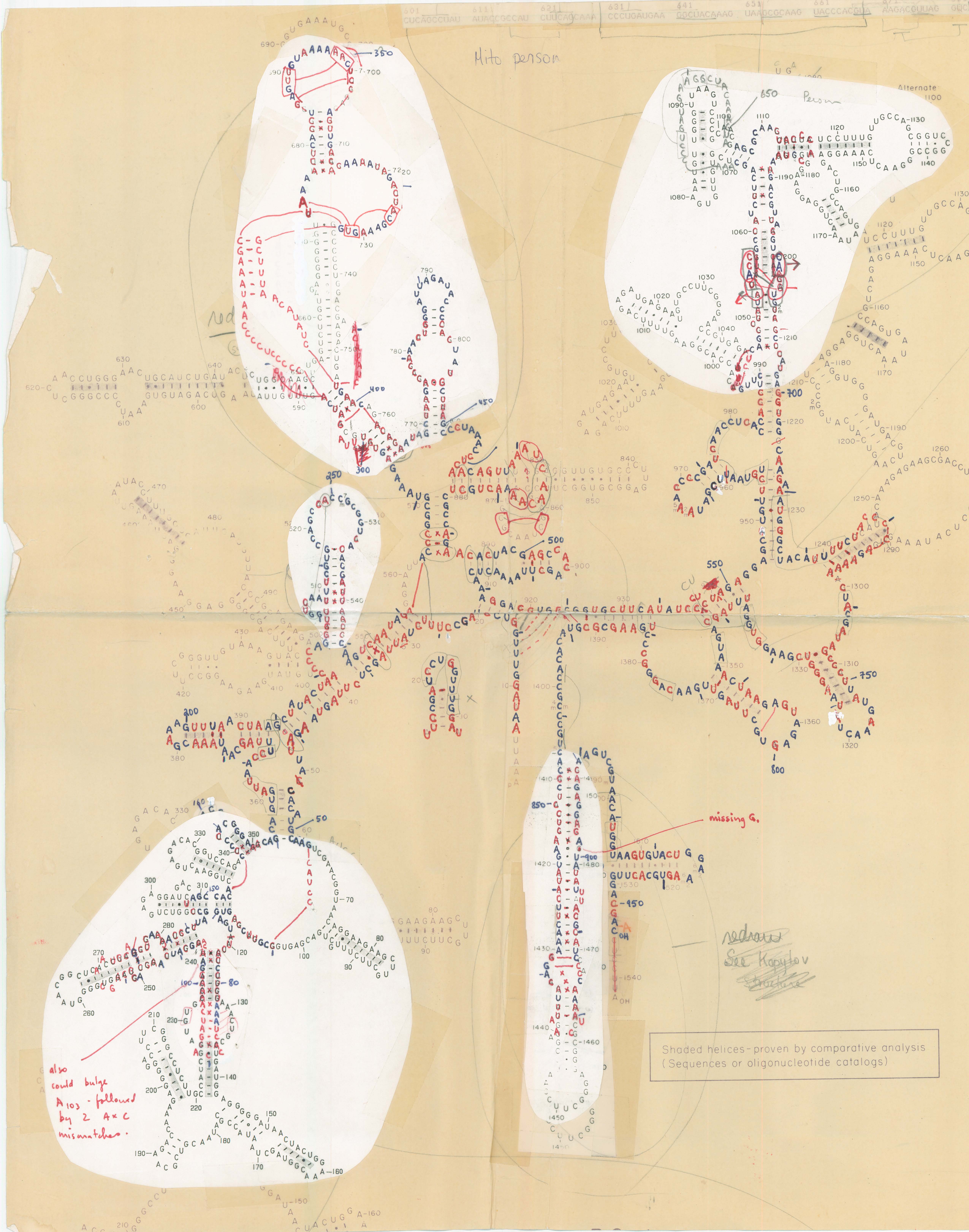
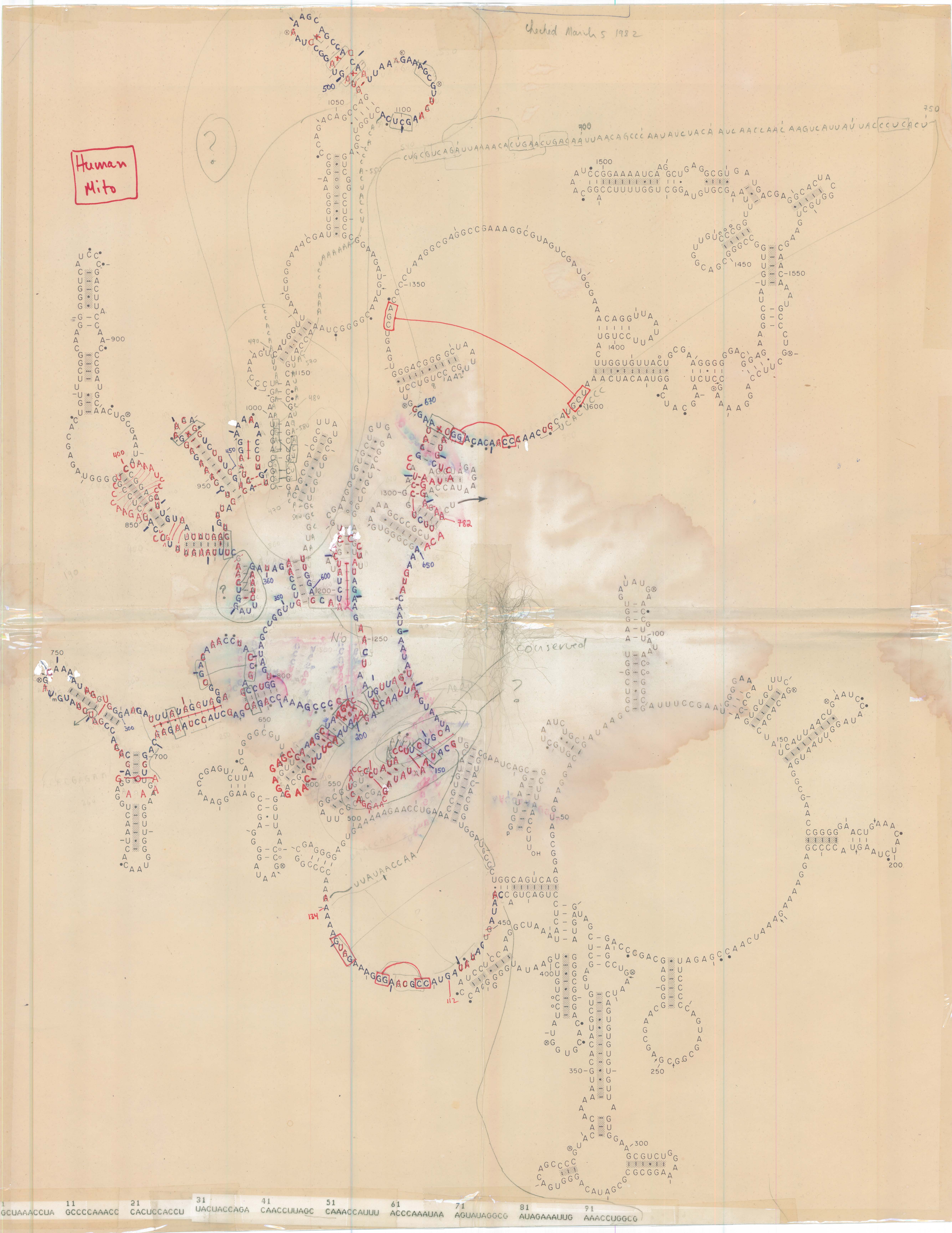
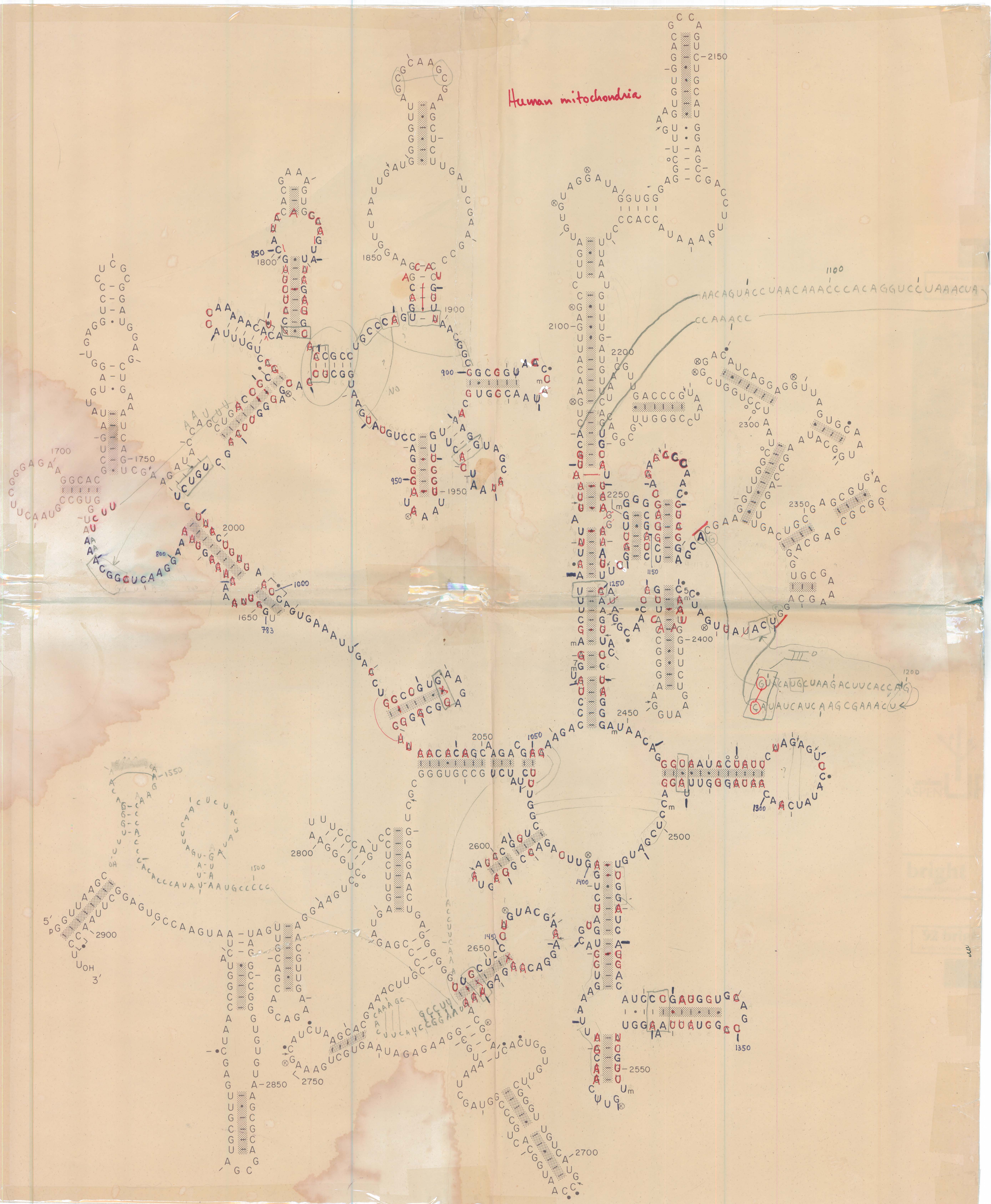
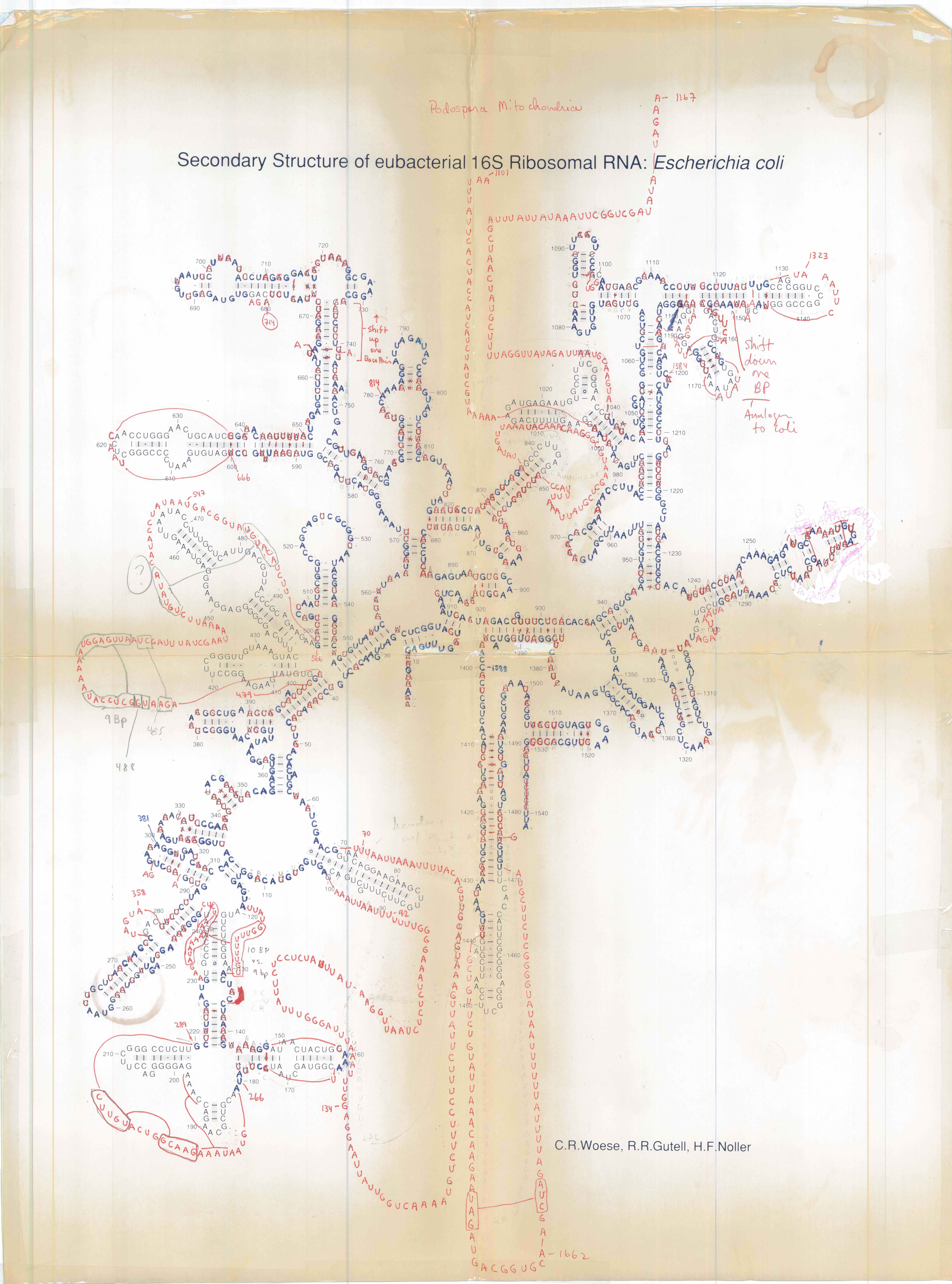
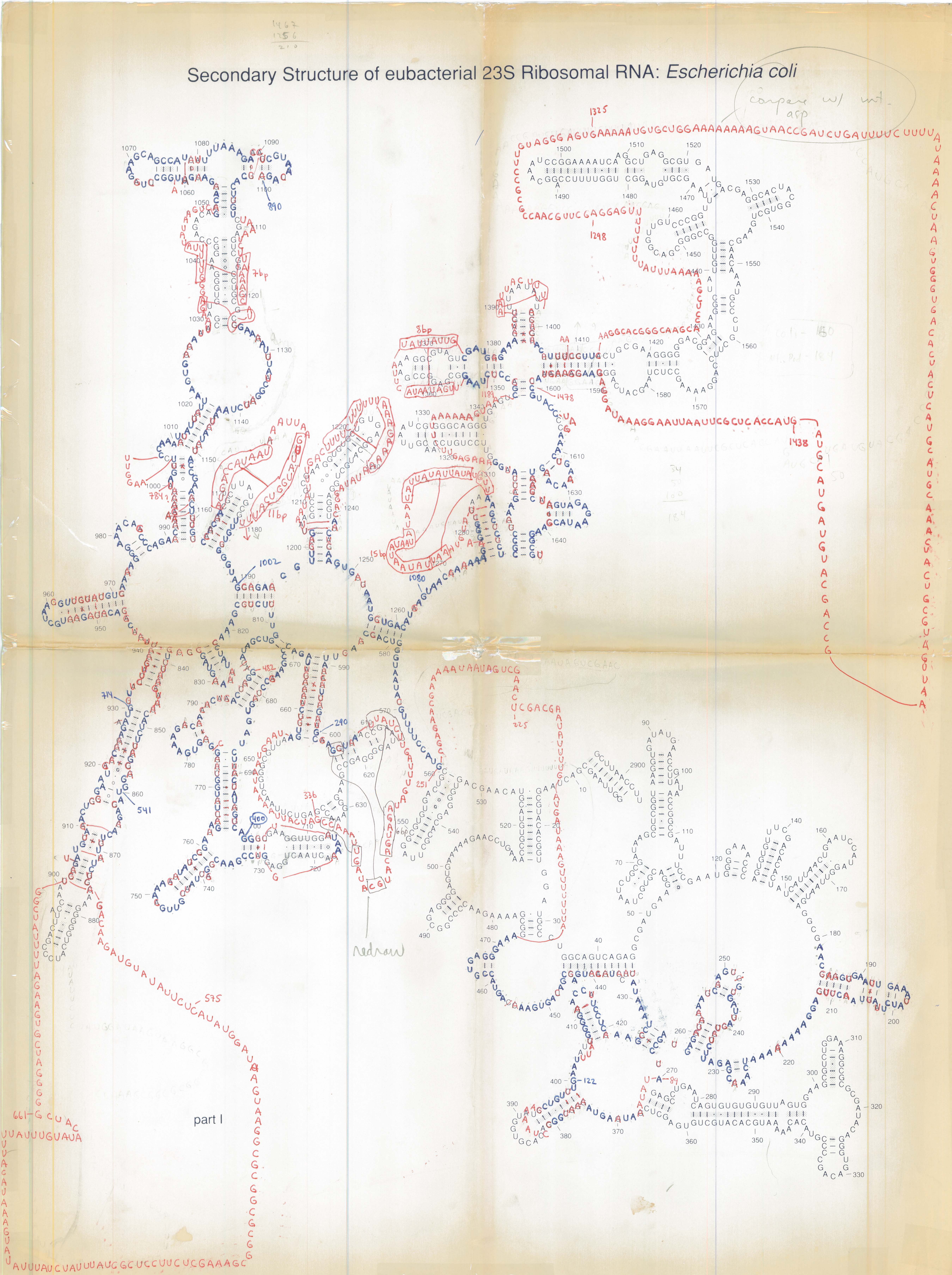
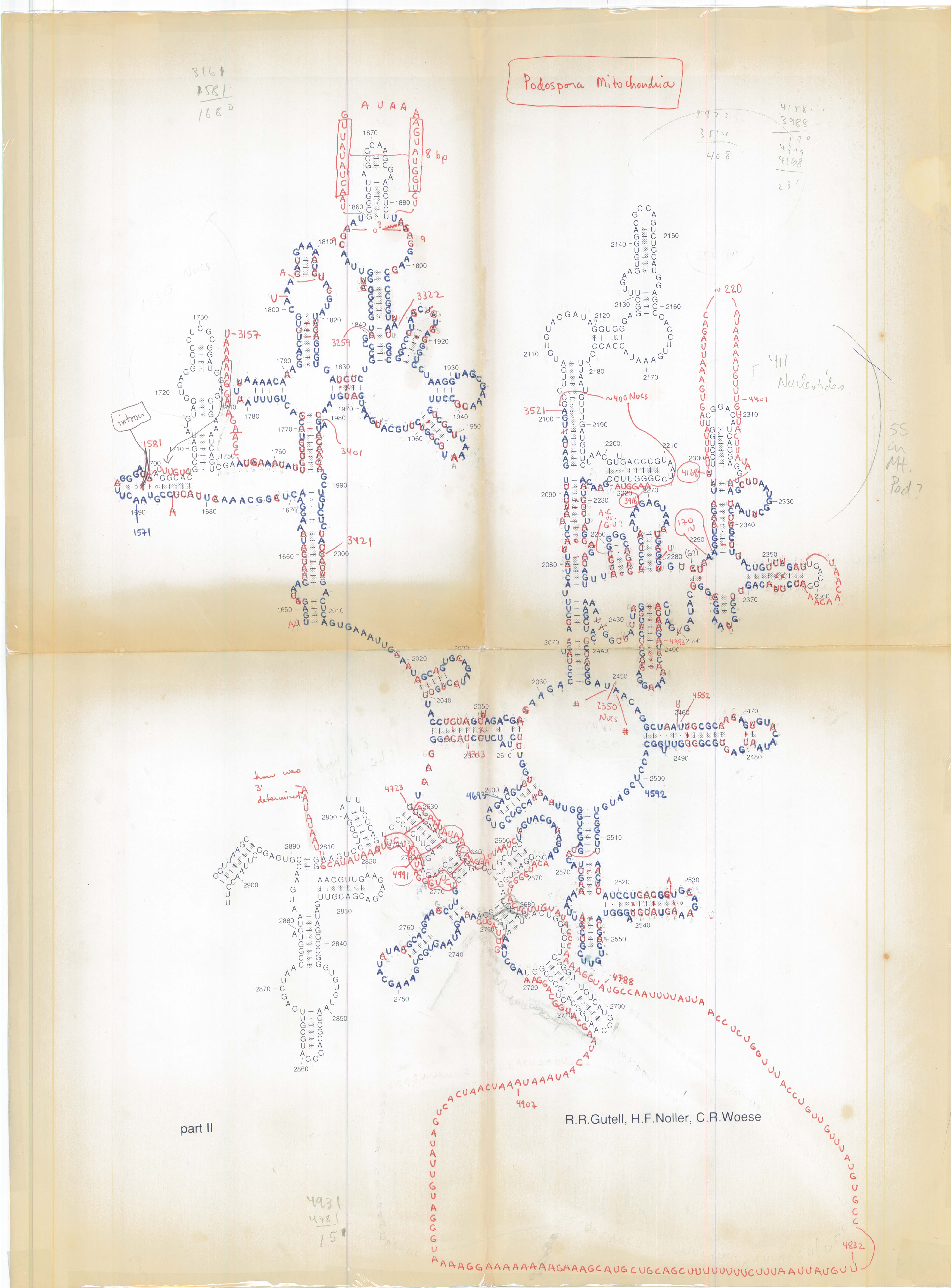
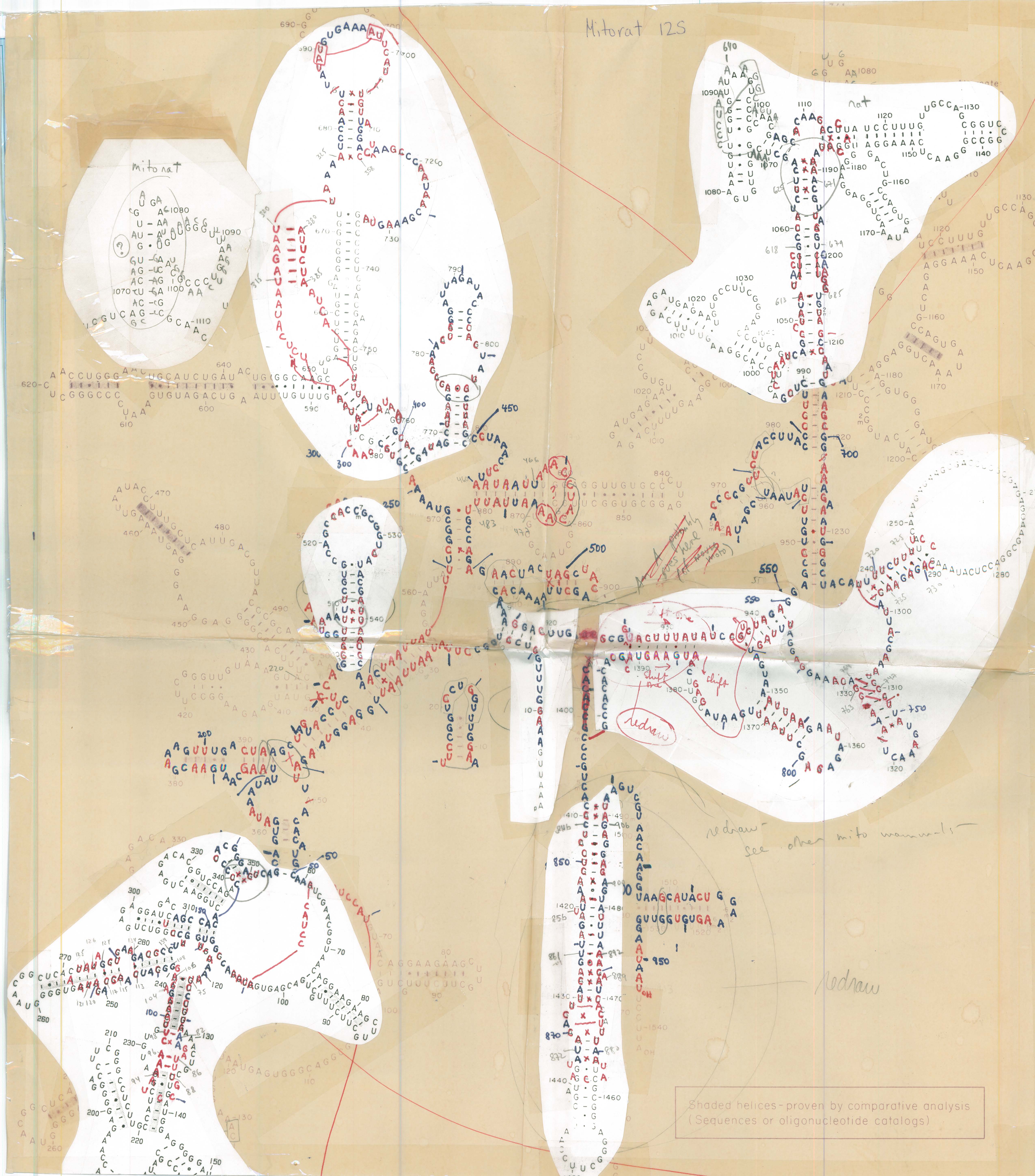
Manual drawing and comparative modeling of RNA sequences in the 1980s was an essential process to determine and refine the 16S (and 16S-like) and 23S (and 23S-like) rRNA secondary structures [1-3]. These efforts were also the foundation for the:
- determination of rRNA structure evolution, and characterization of sequence and structure conservation and variation [3-5],
- study of RNA structural motifs and higher-order structure, including pseudoknots and tetraloops [6-9], and
- process of structure-based alignment of RNA sequences [4, 5].
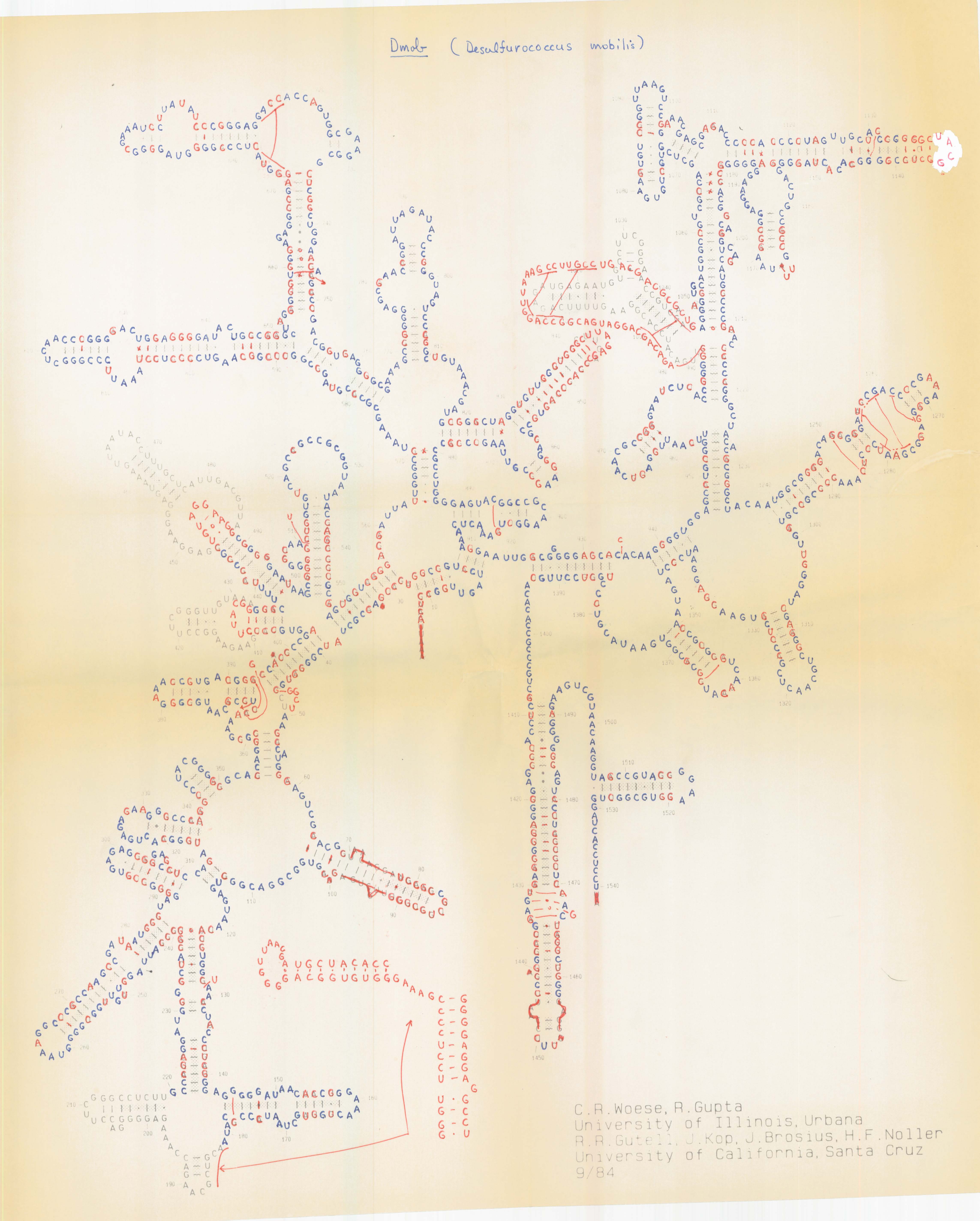
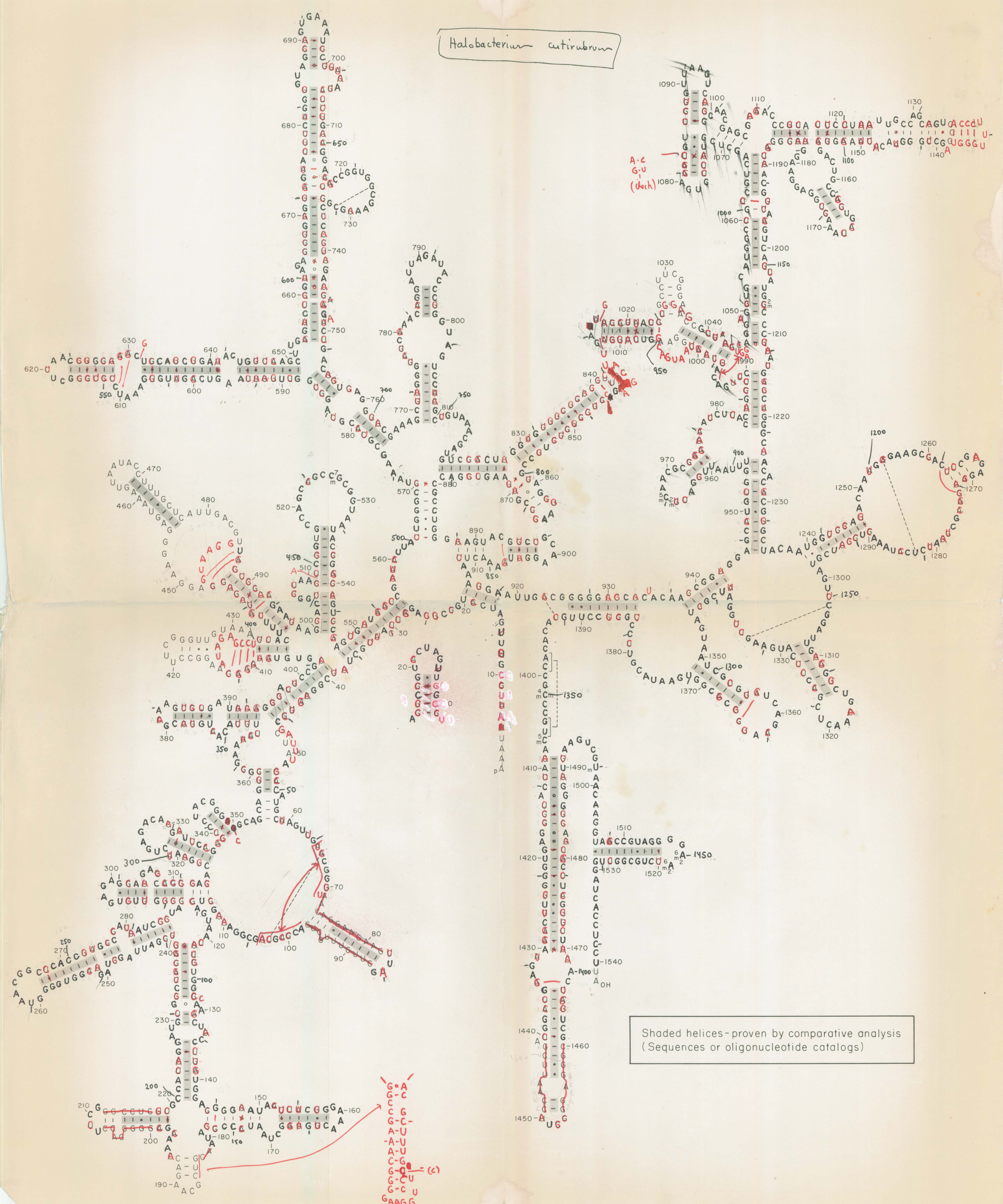
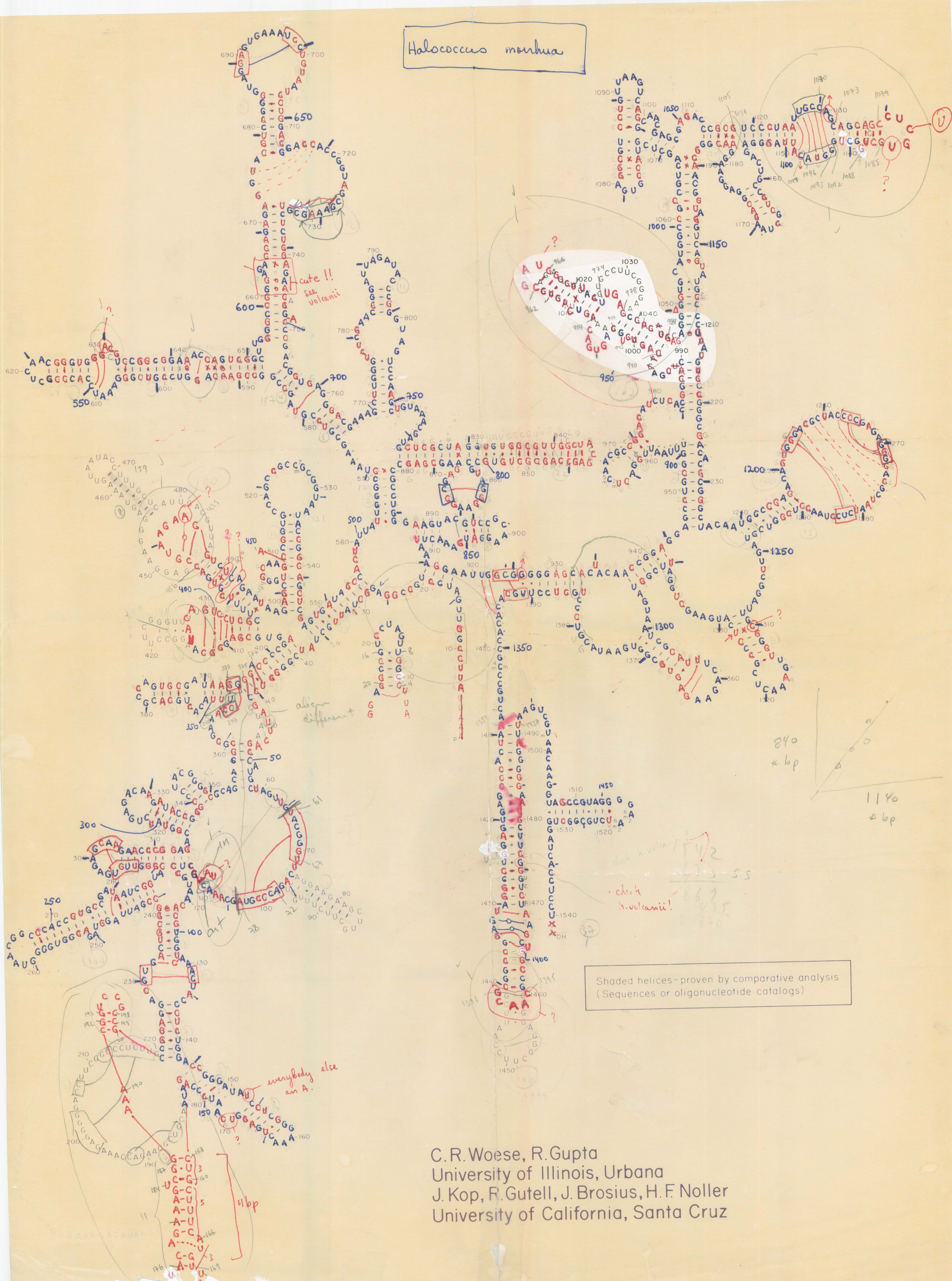
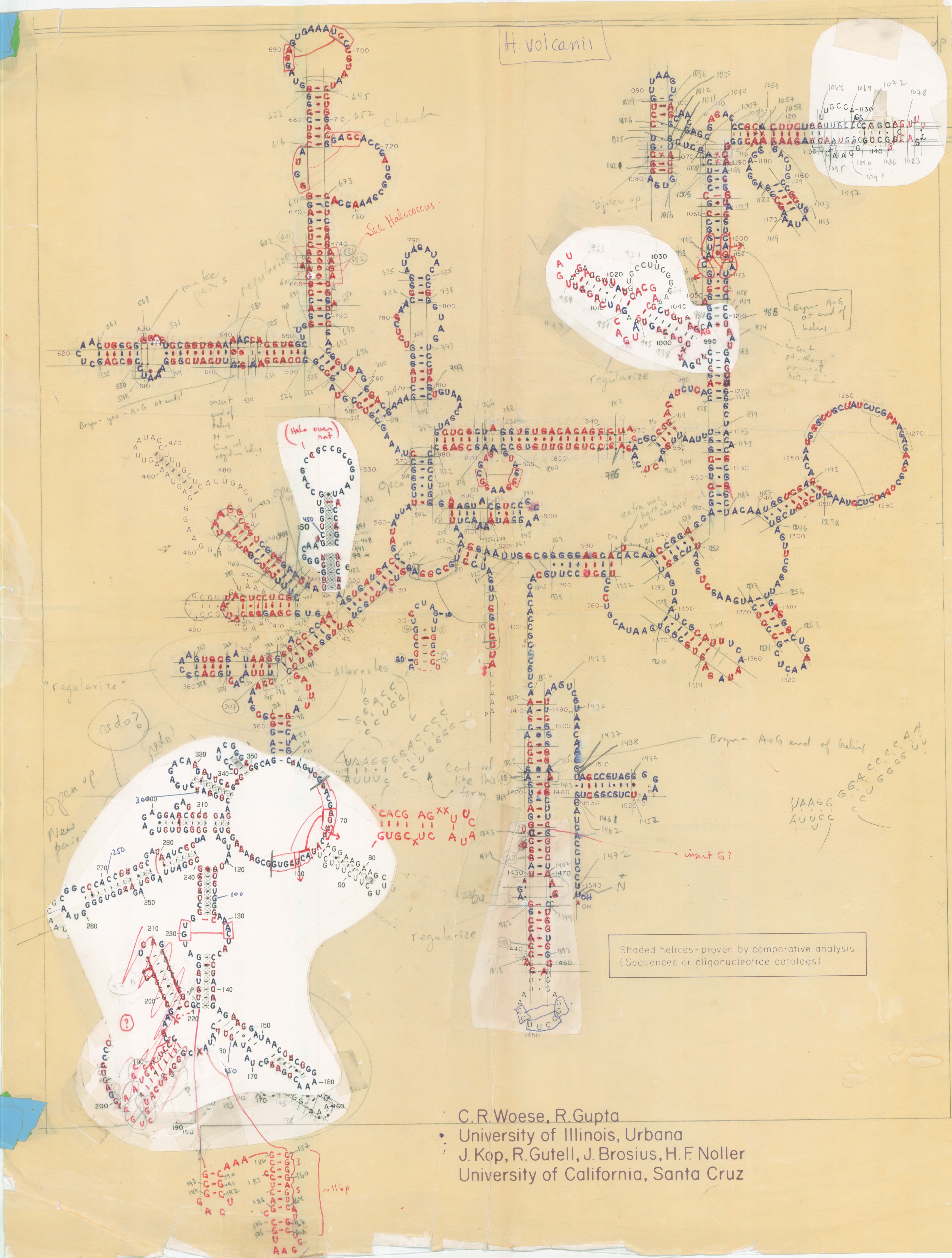
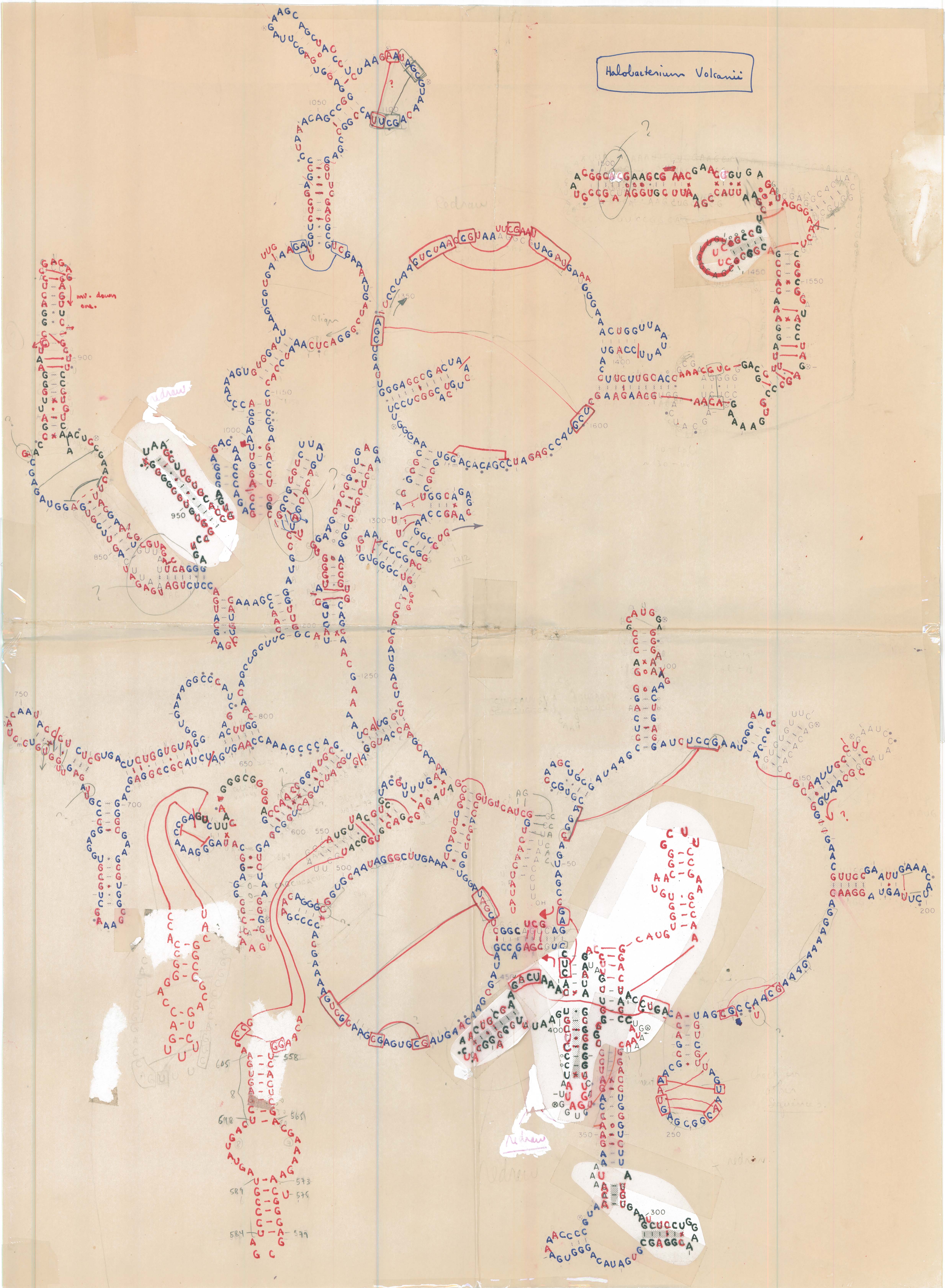
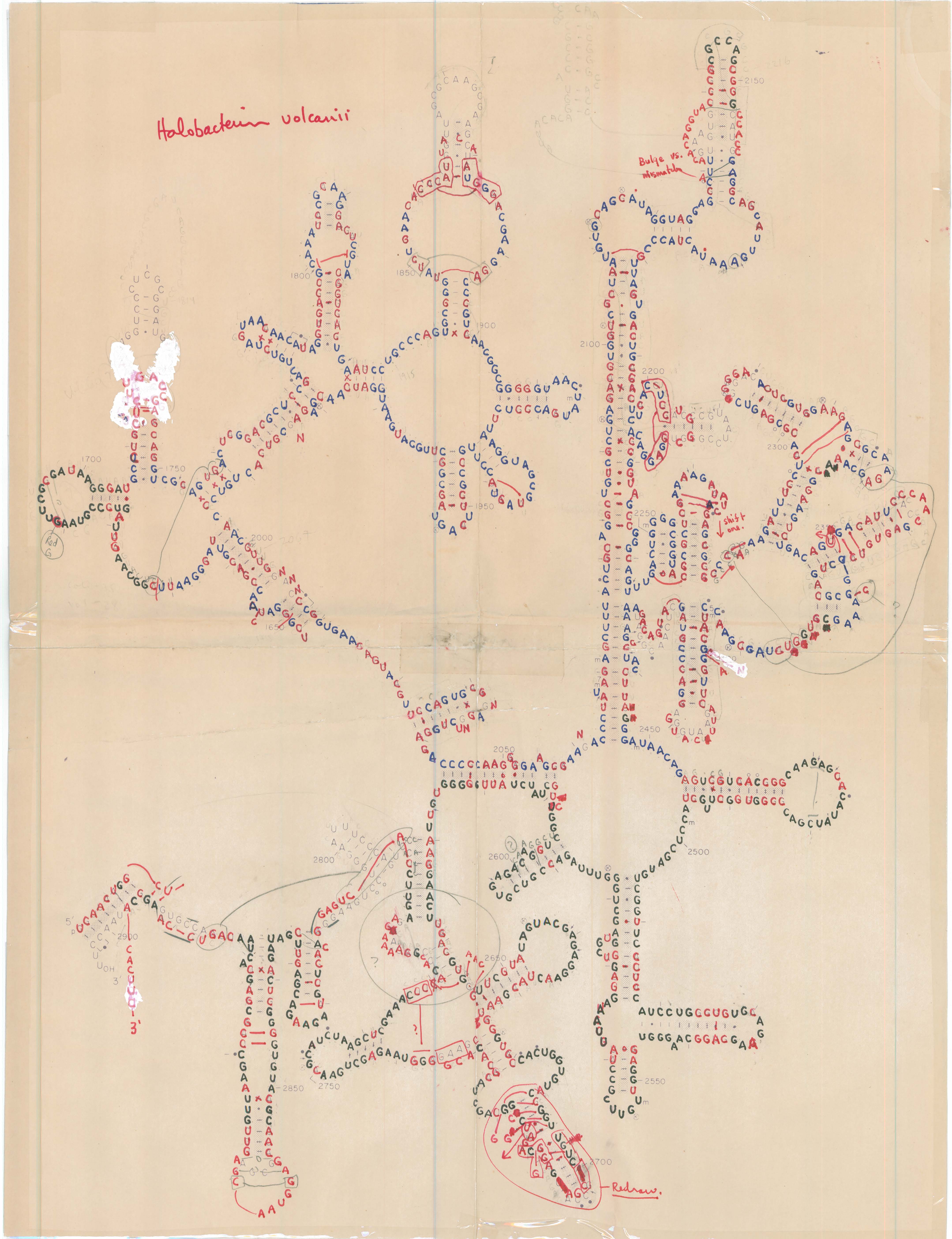
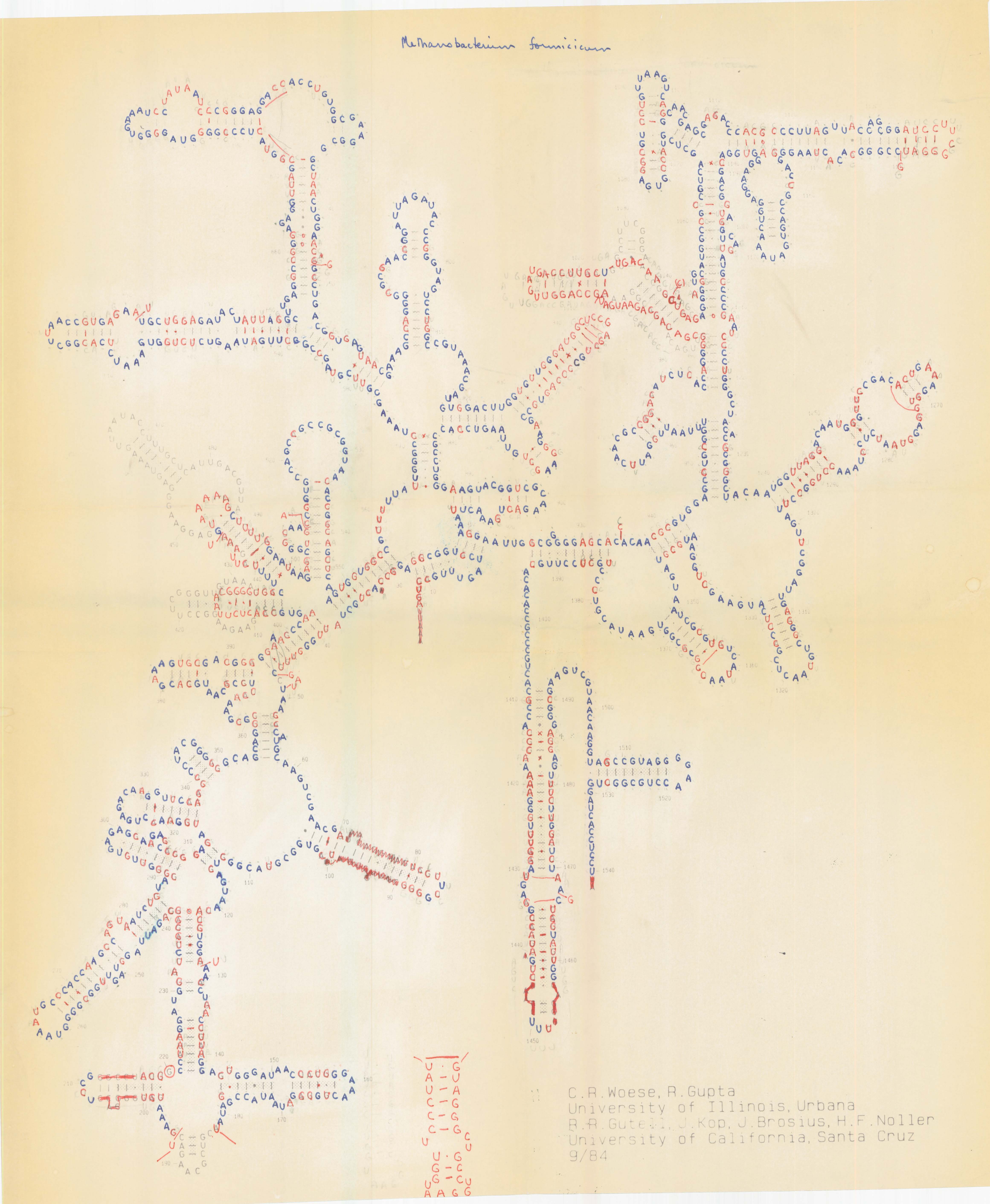
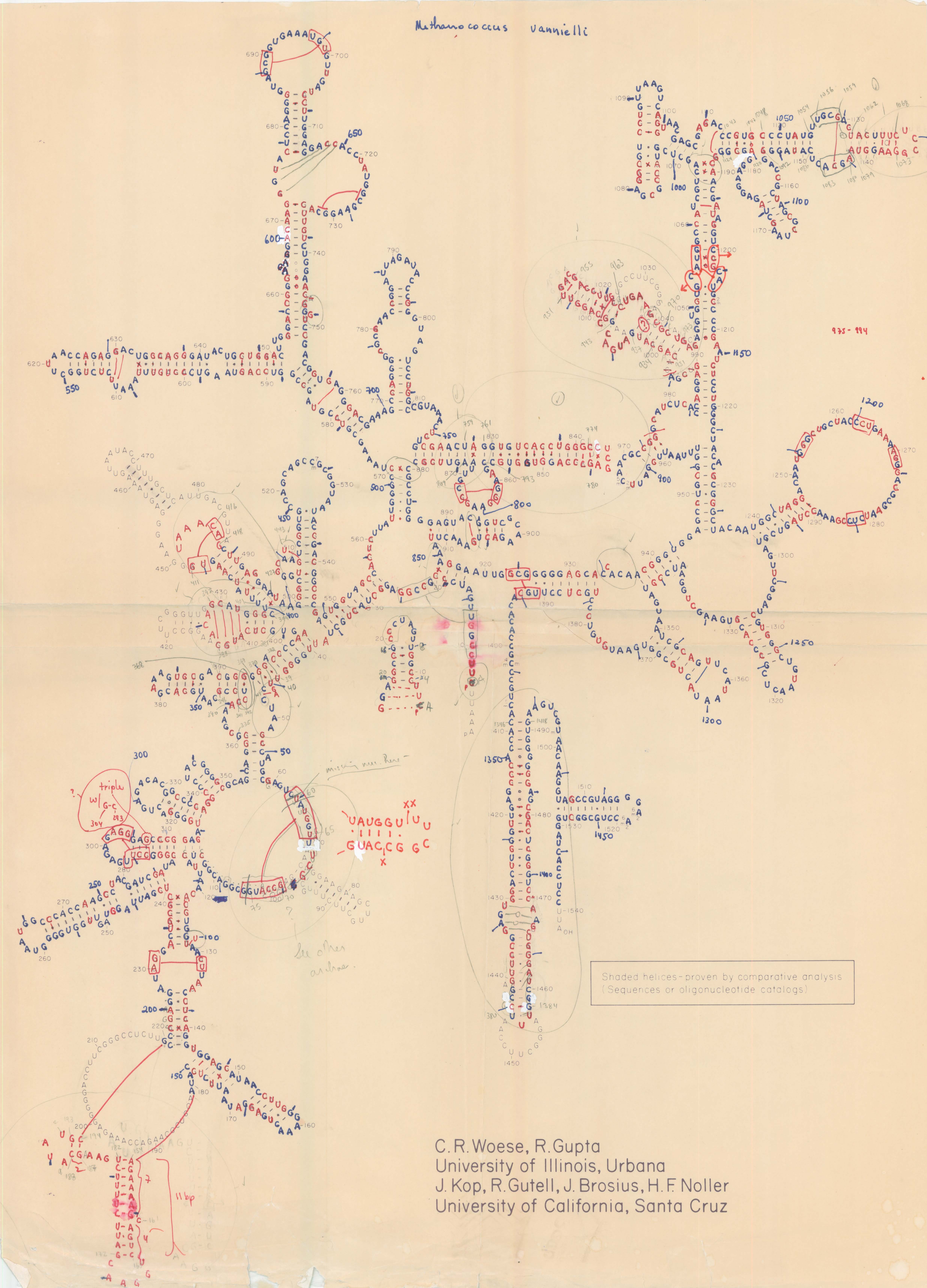
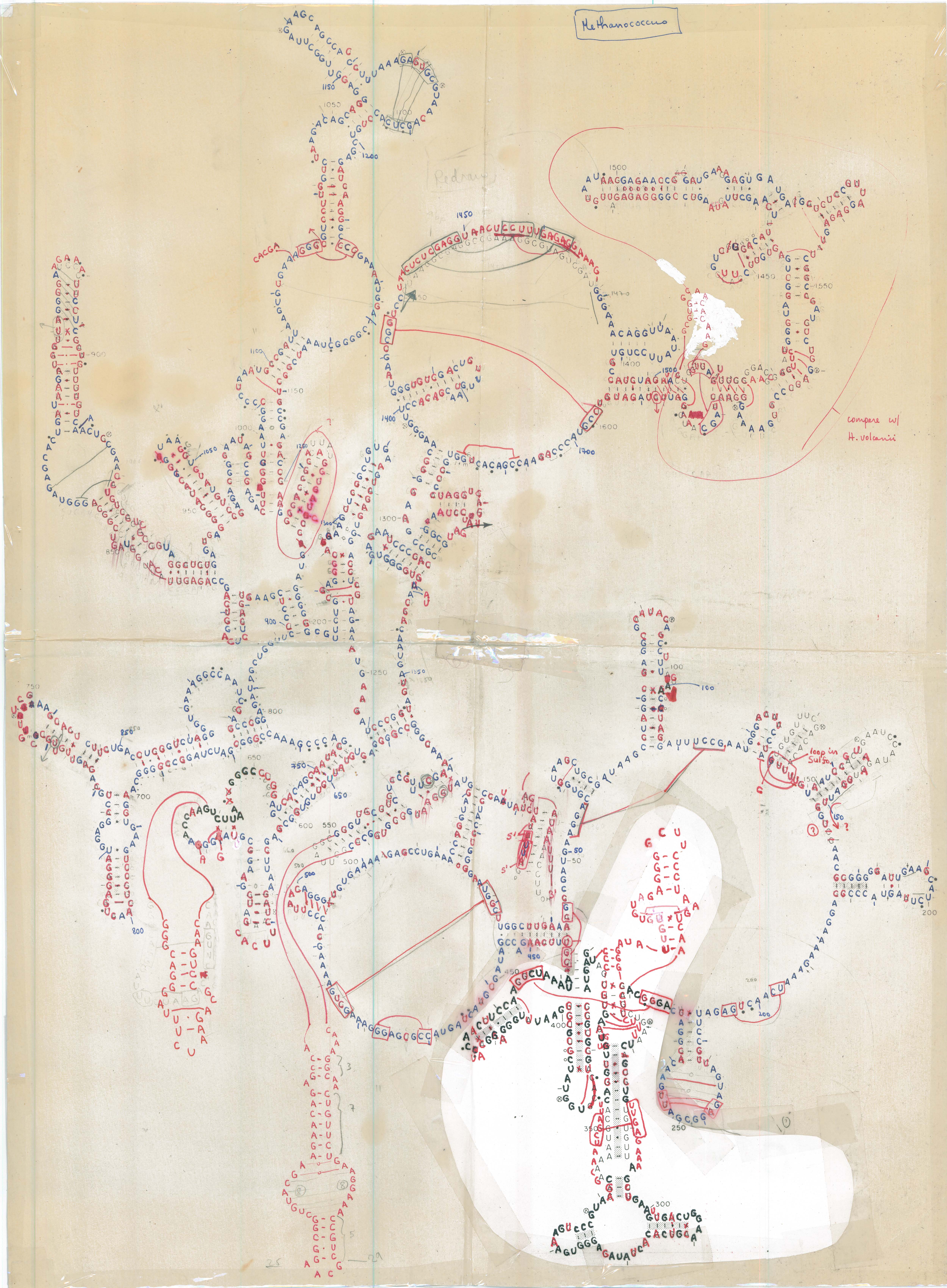
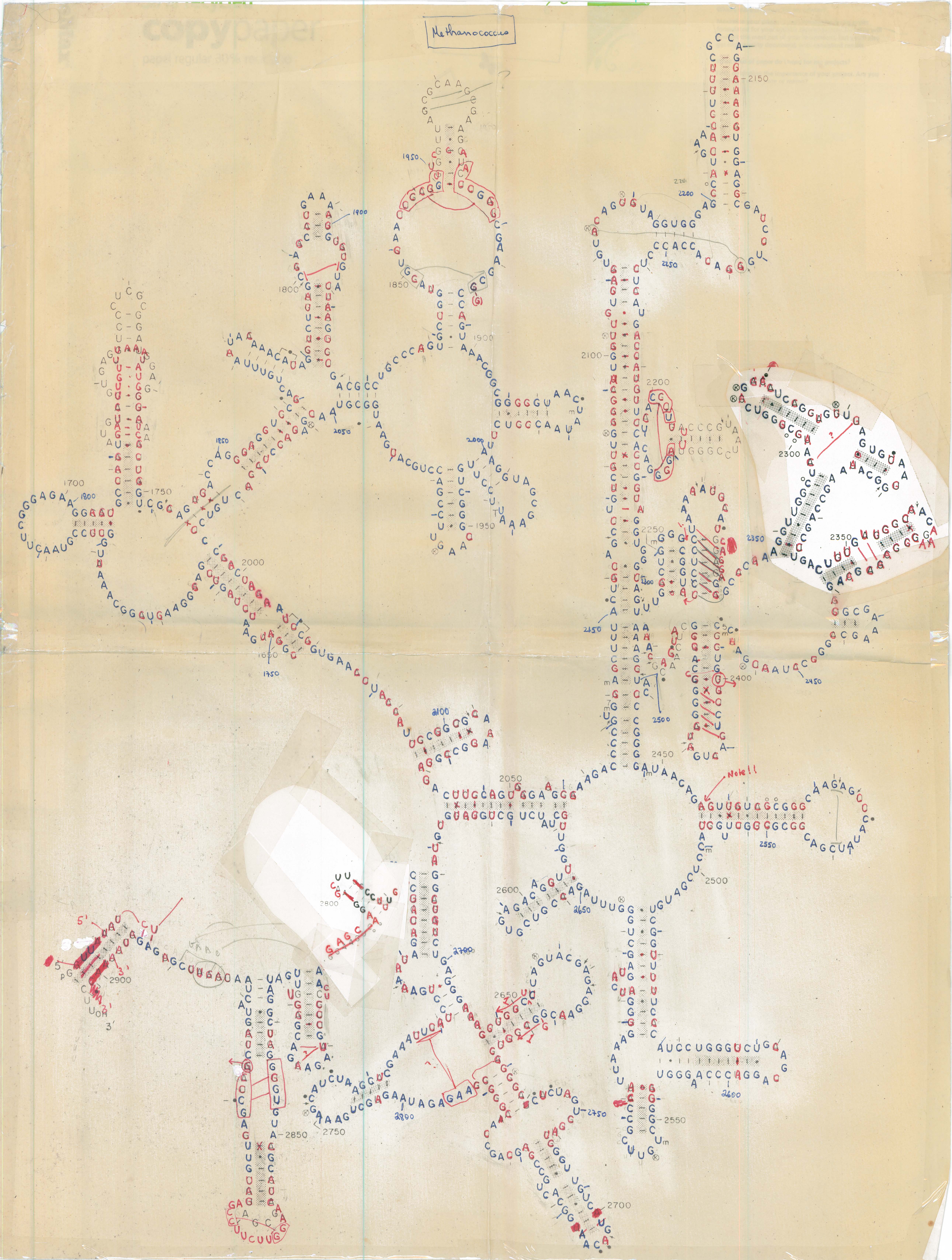
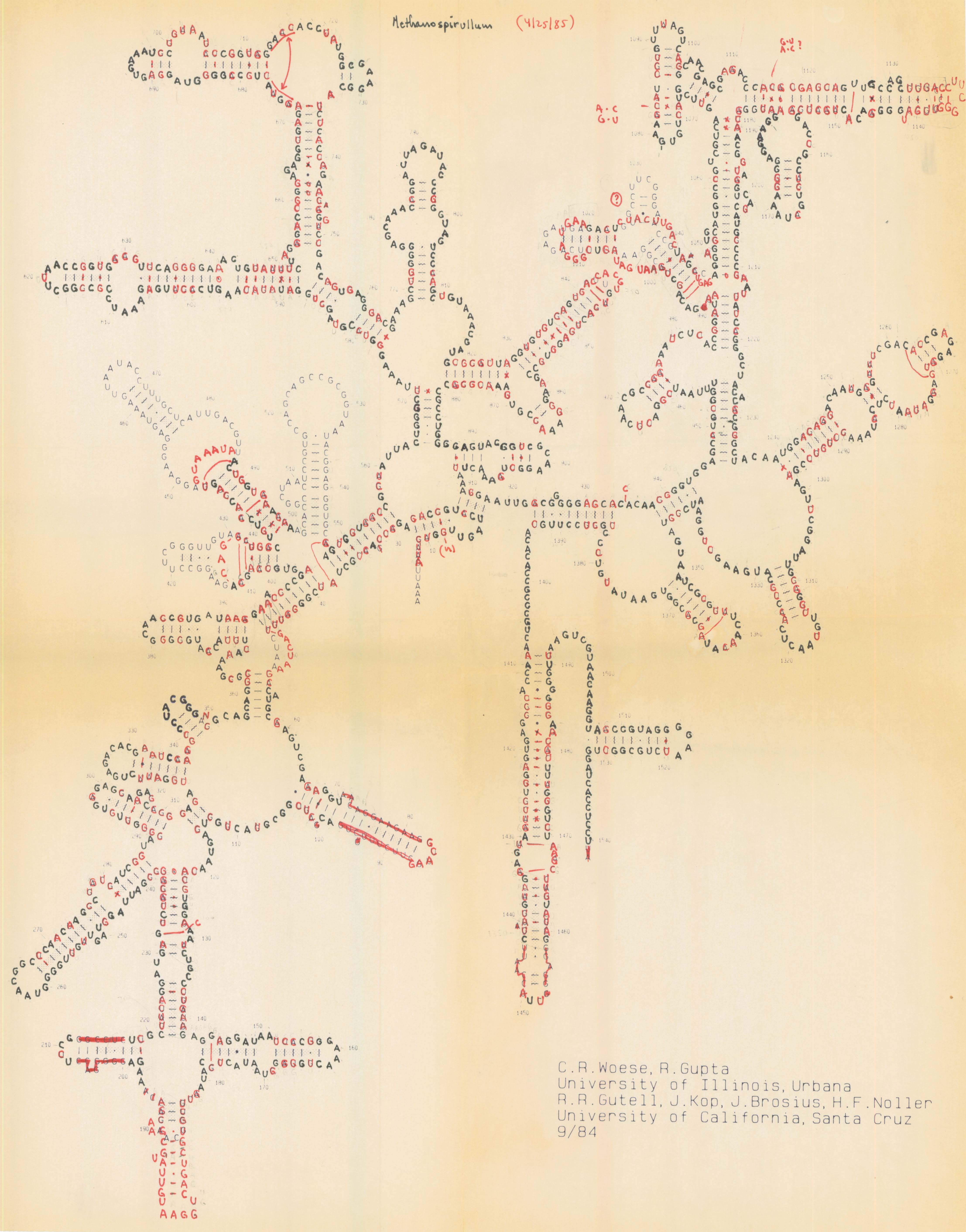
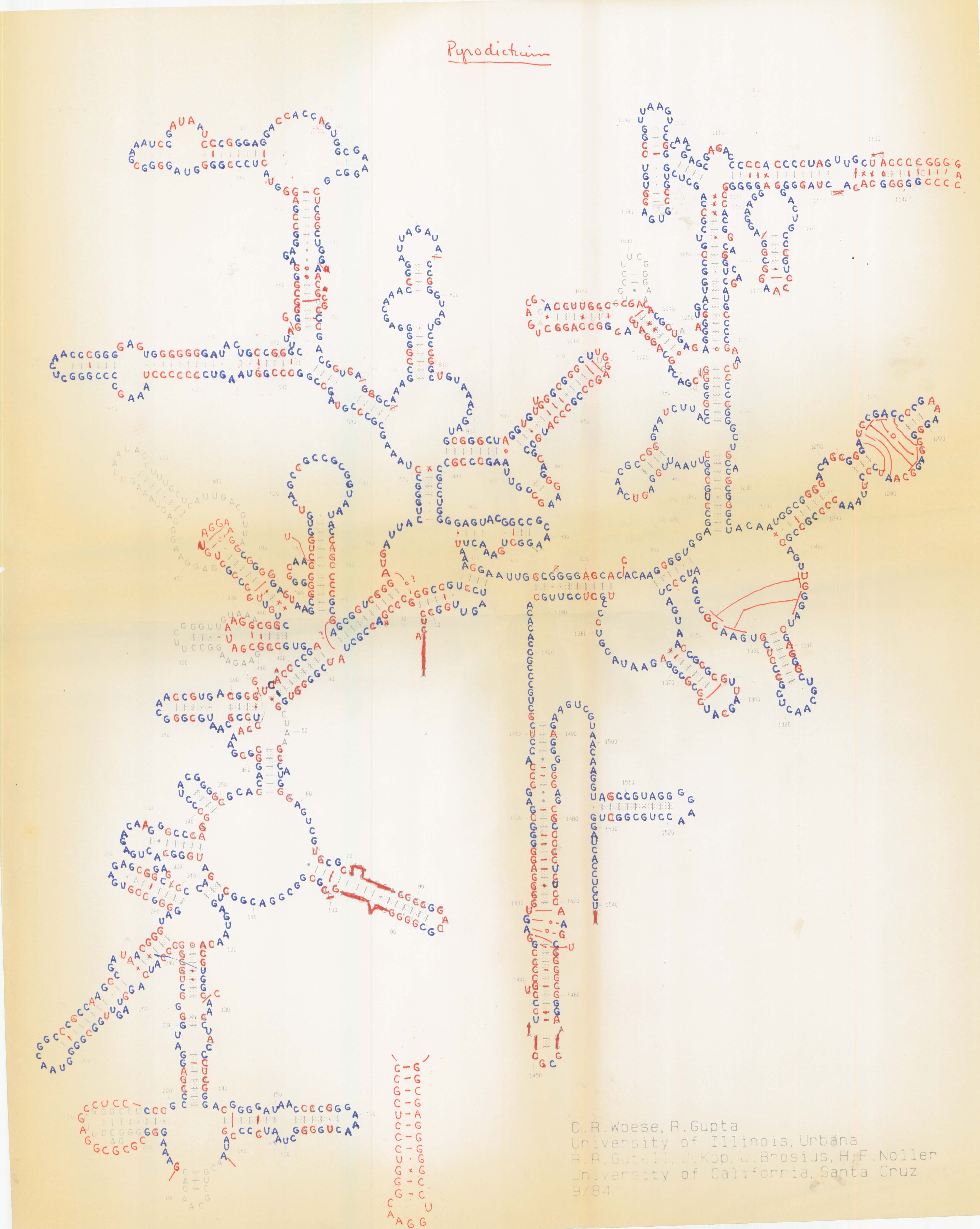
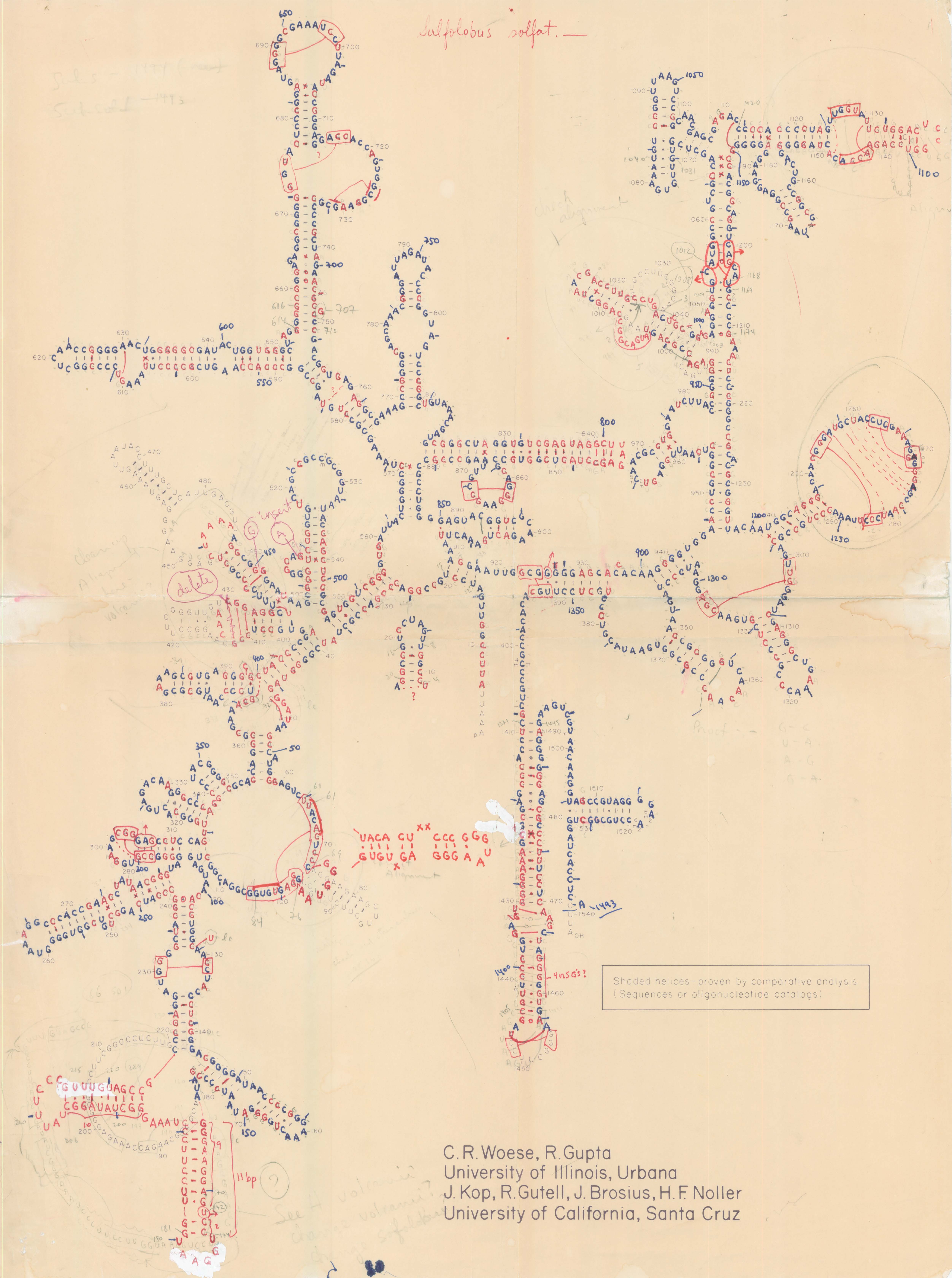
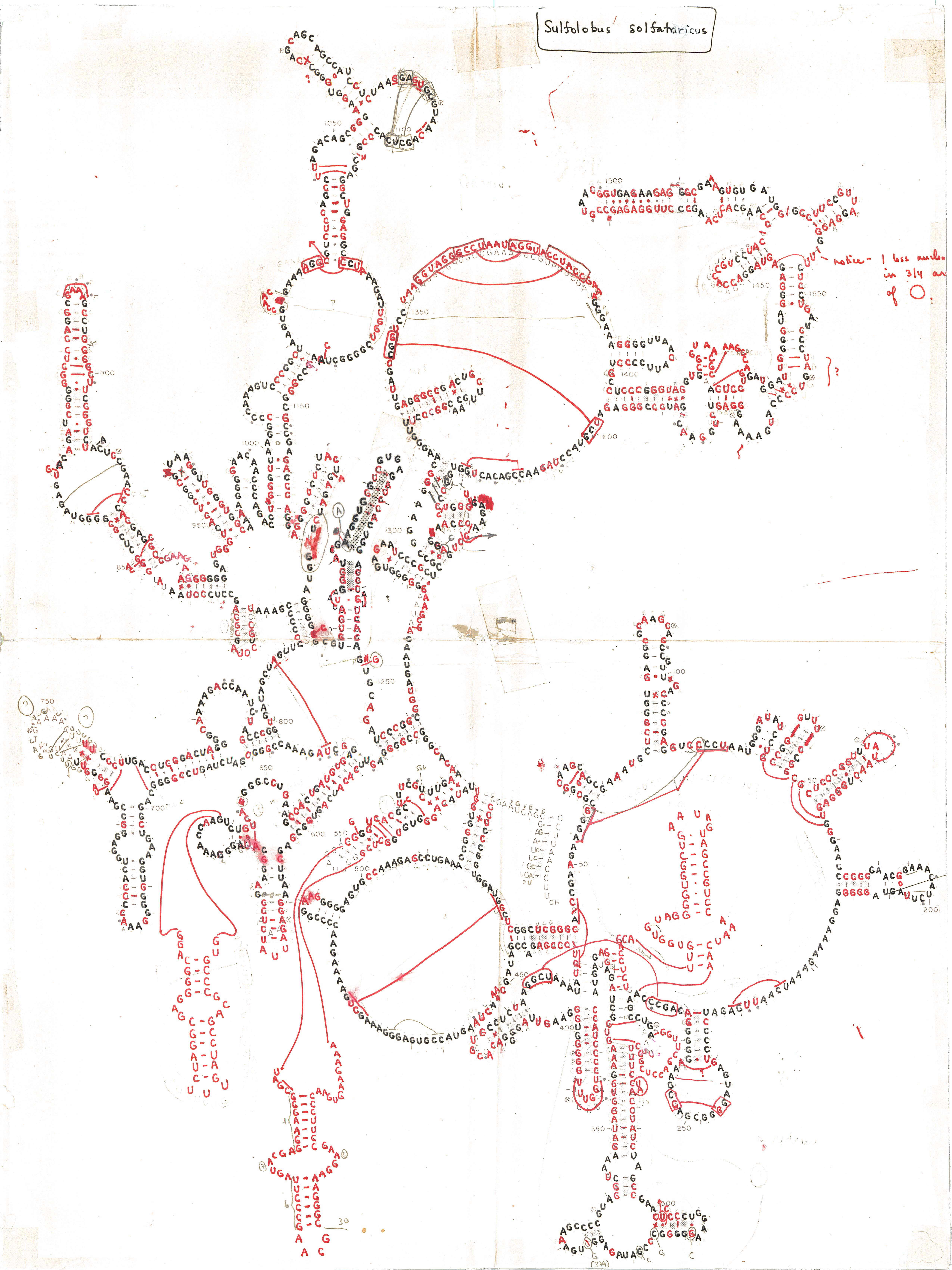
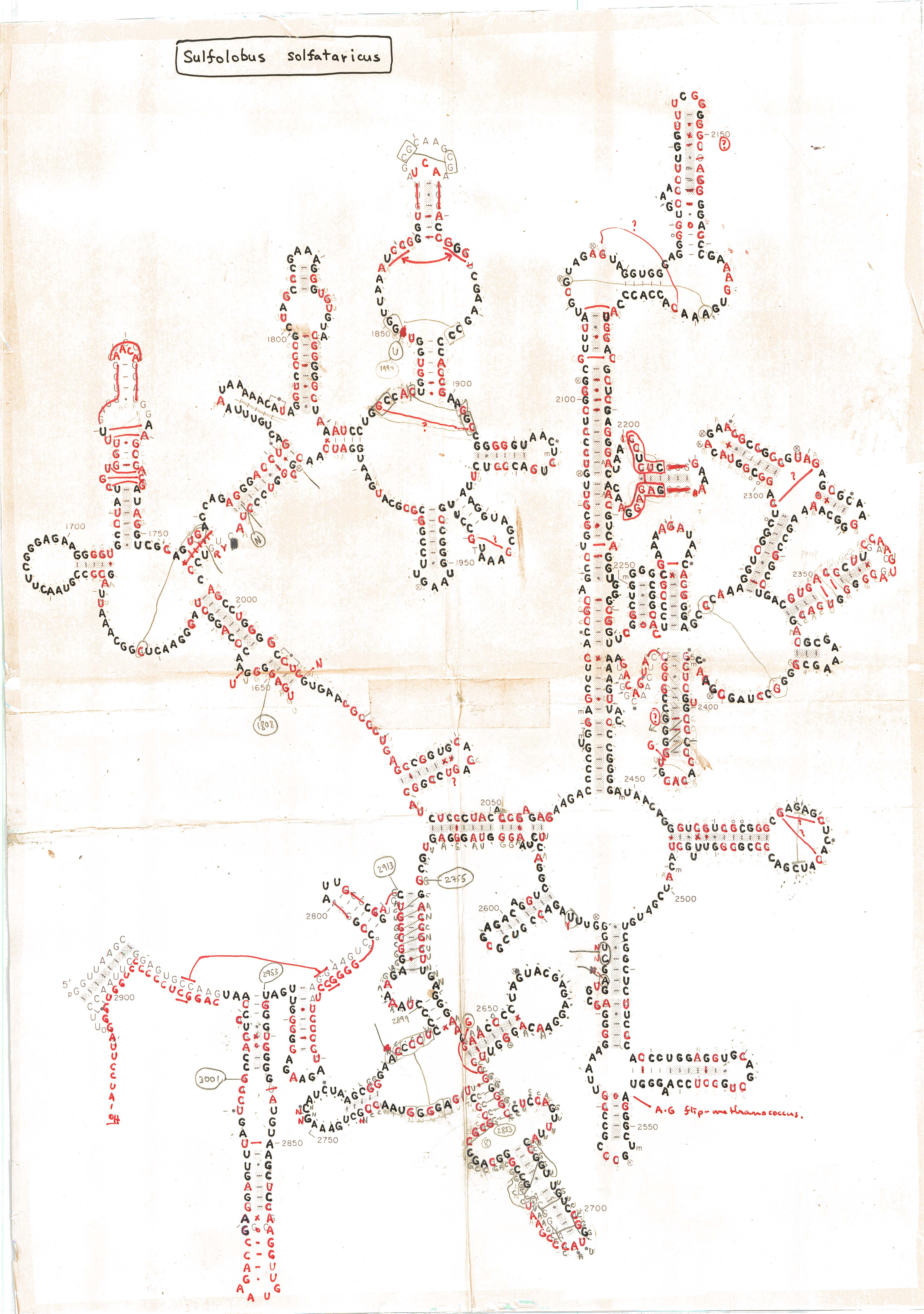
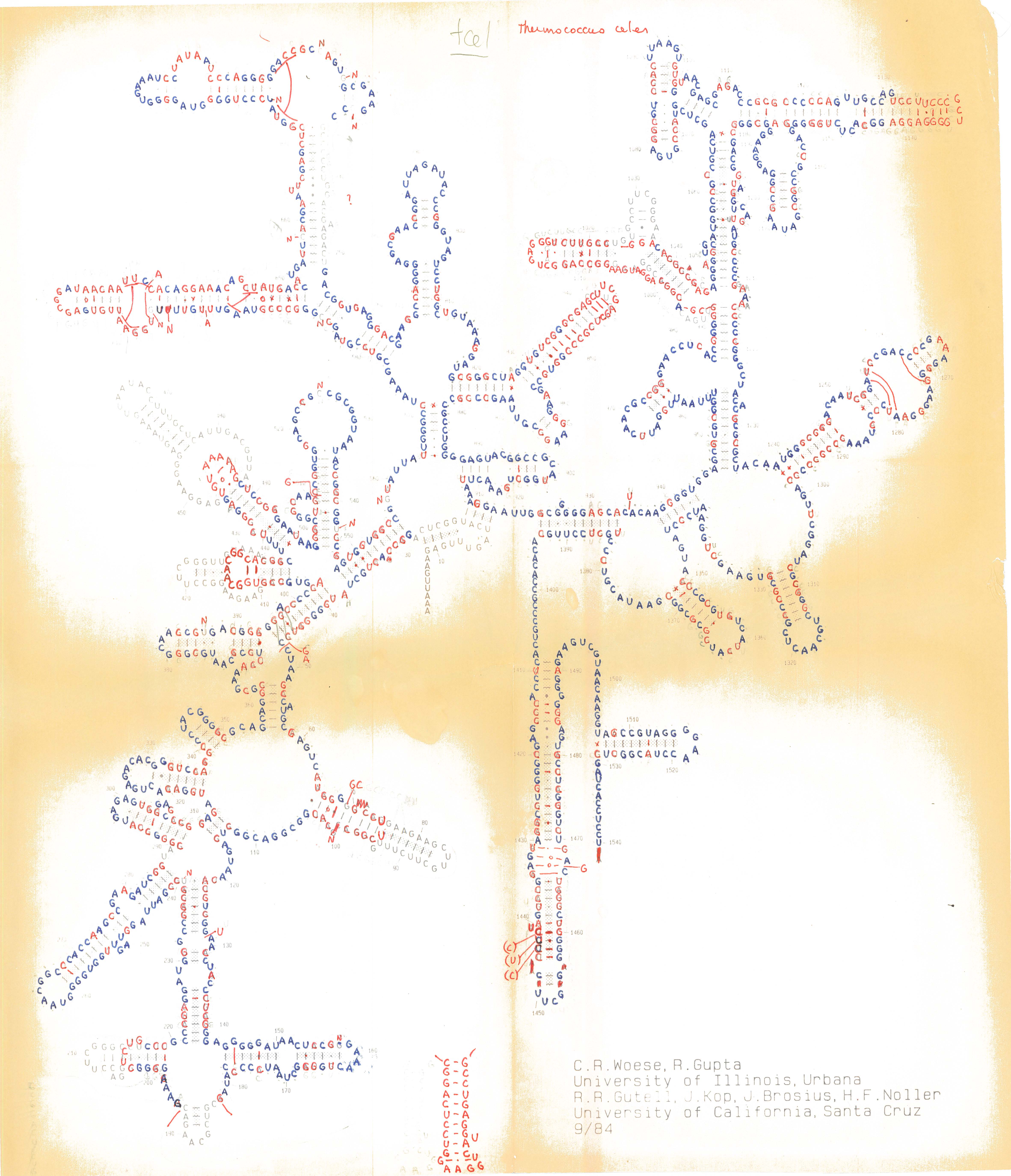
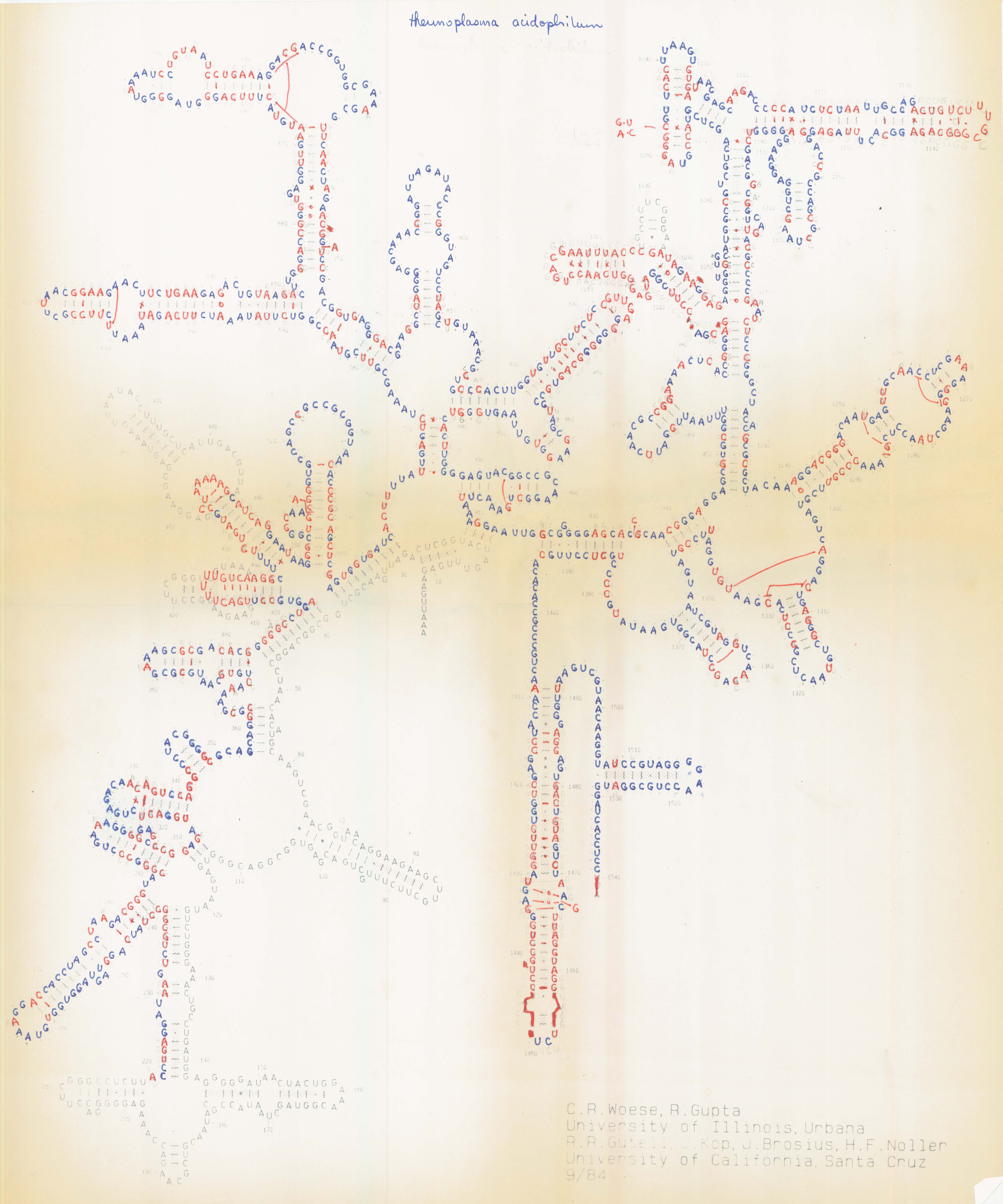
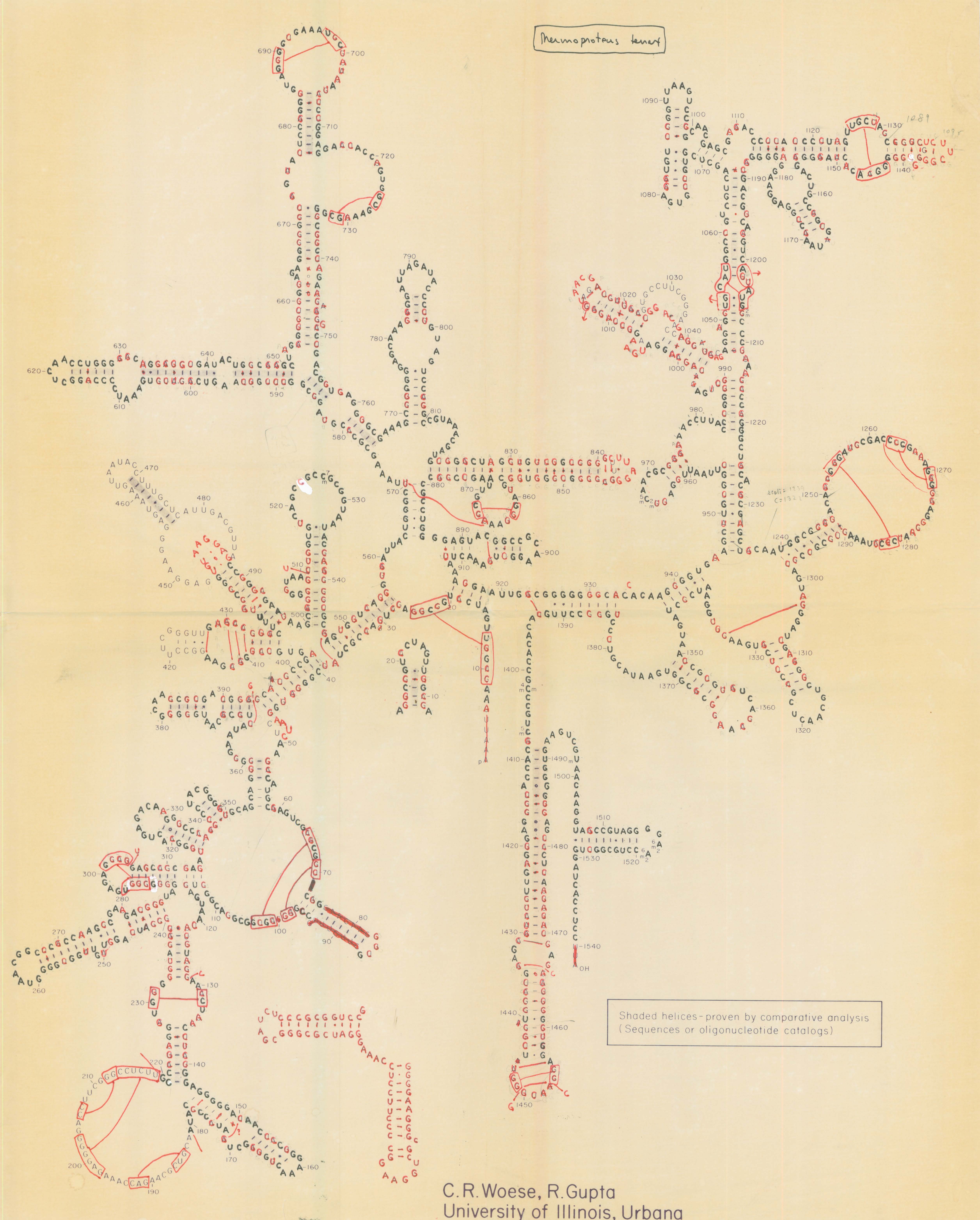
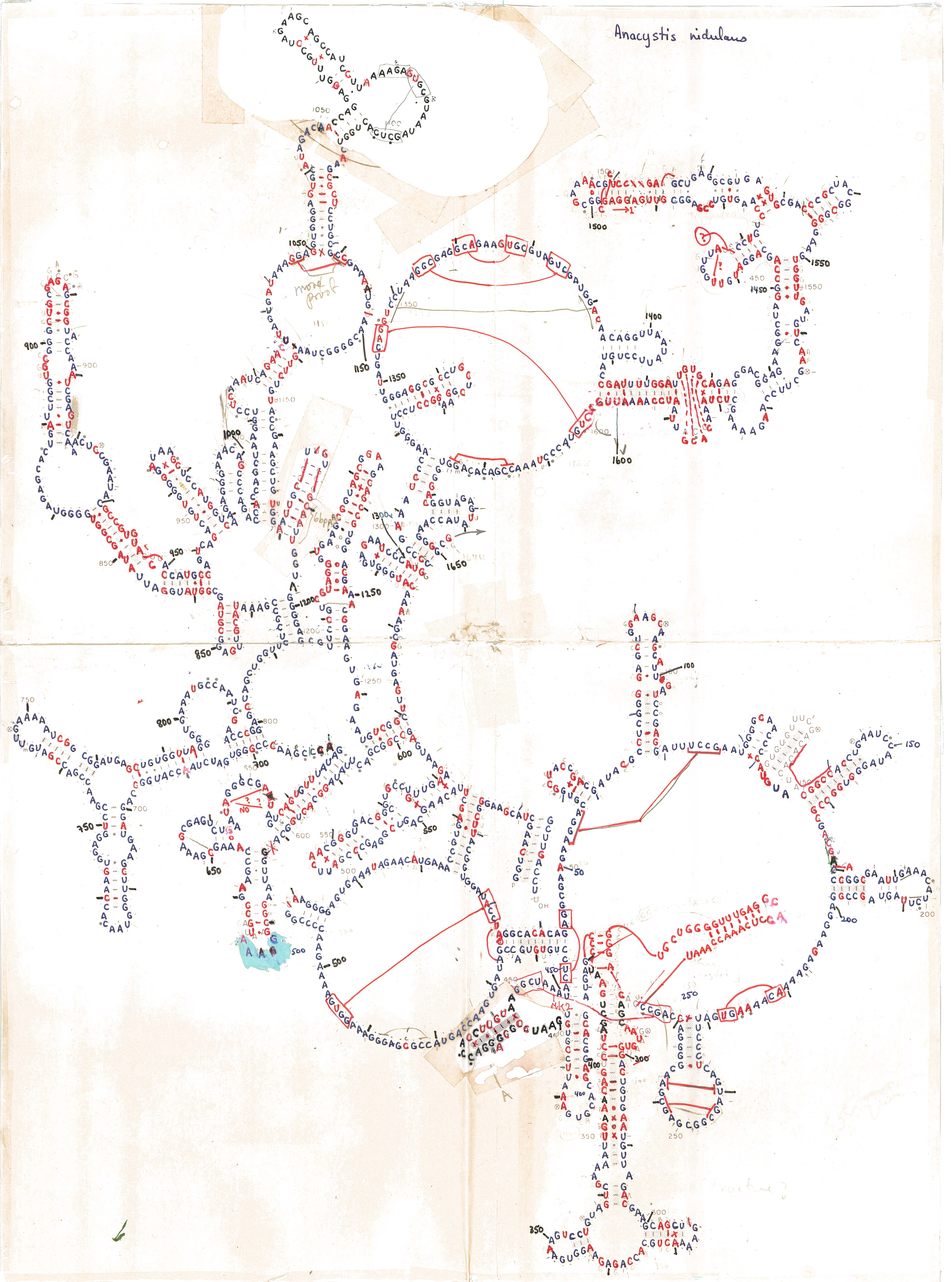
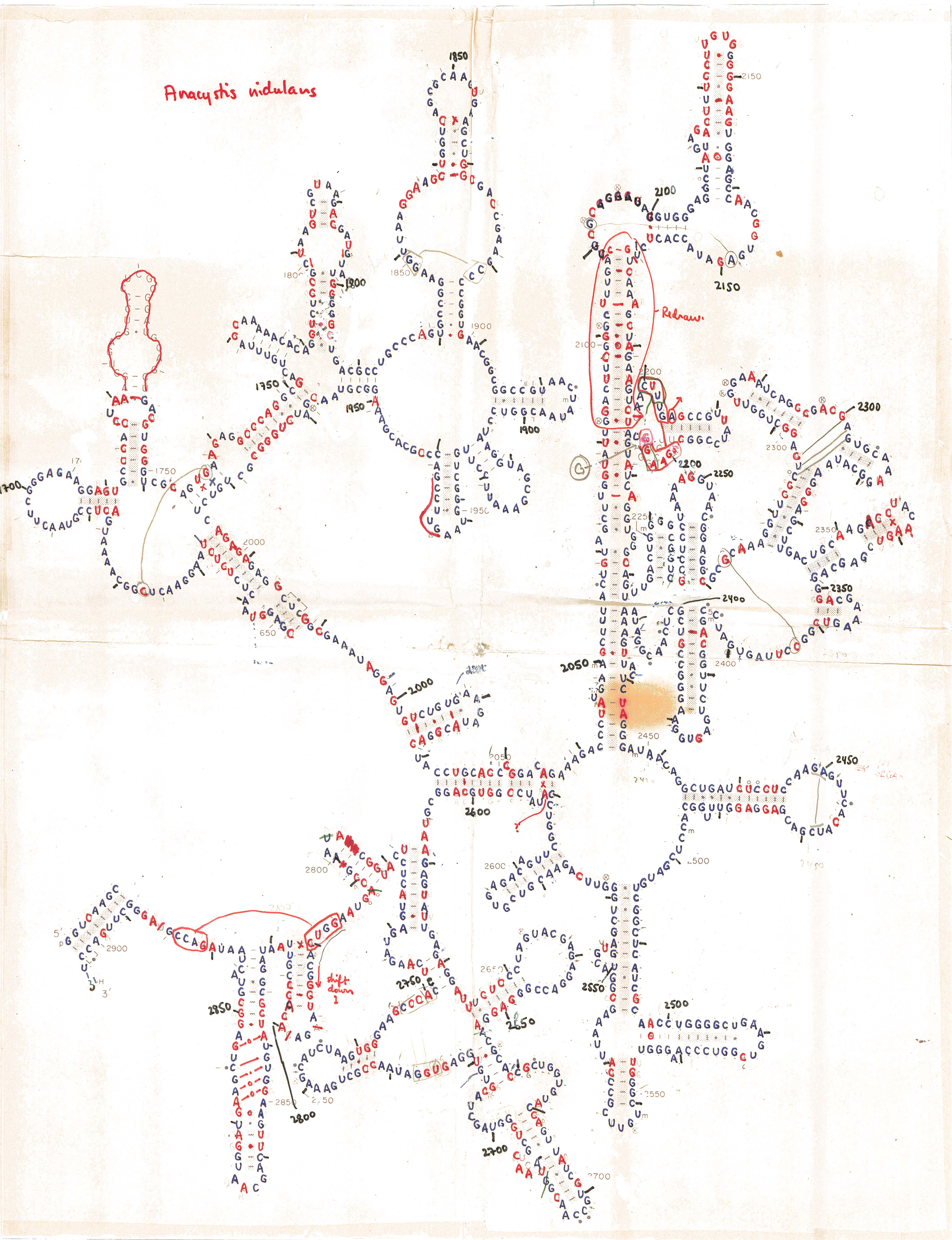
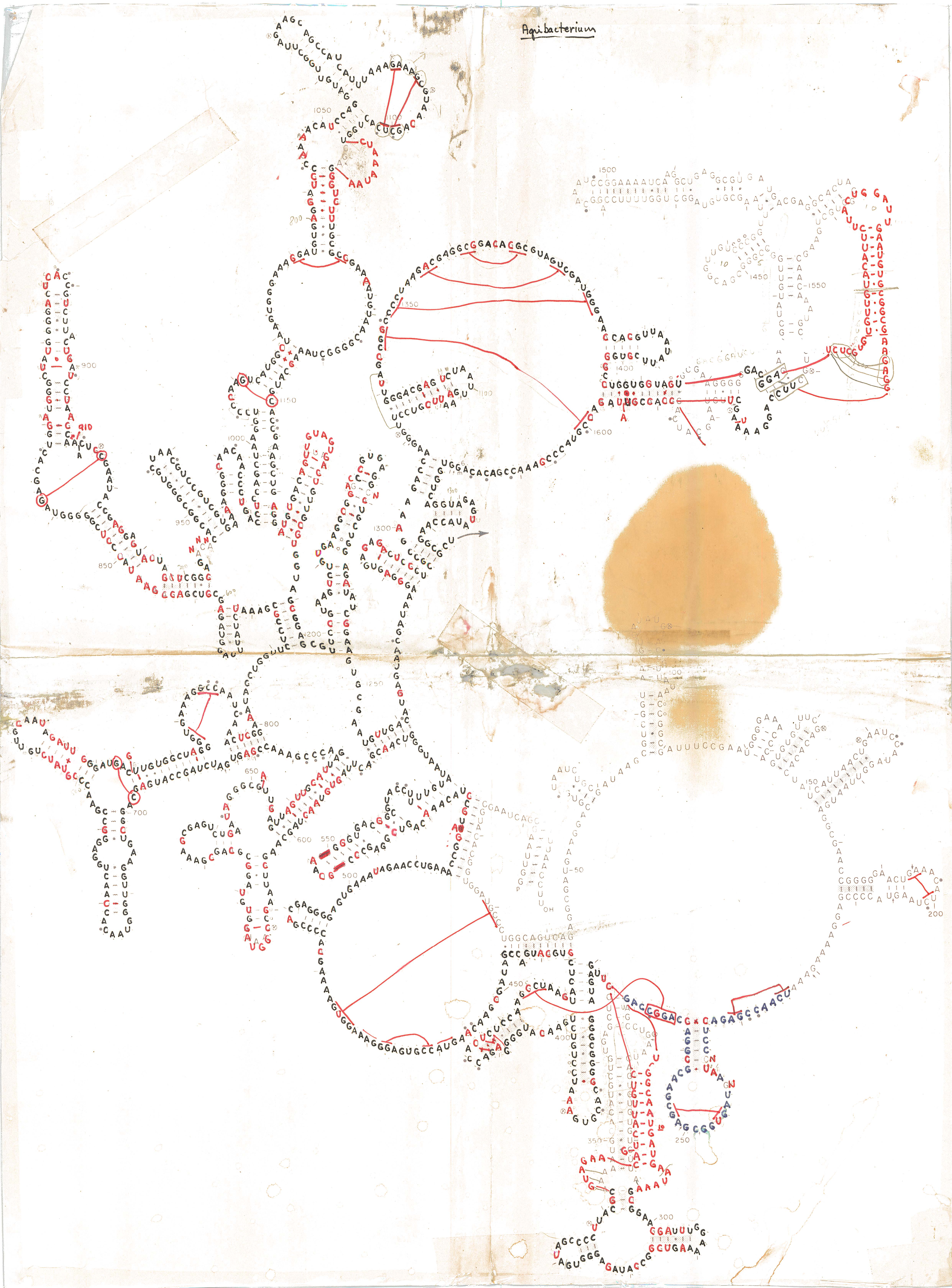
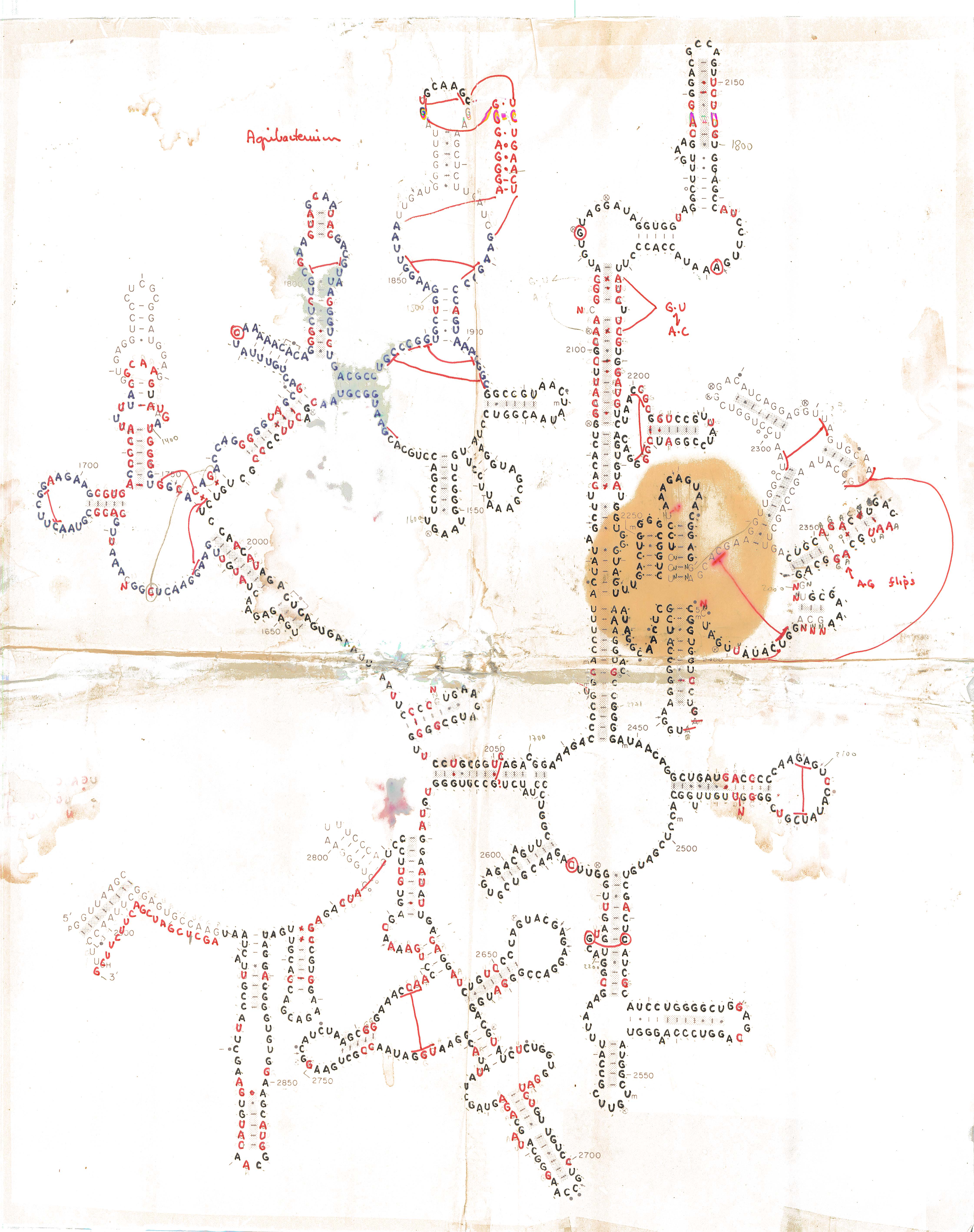
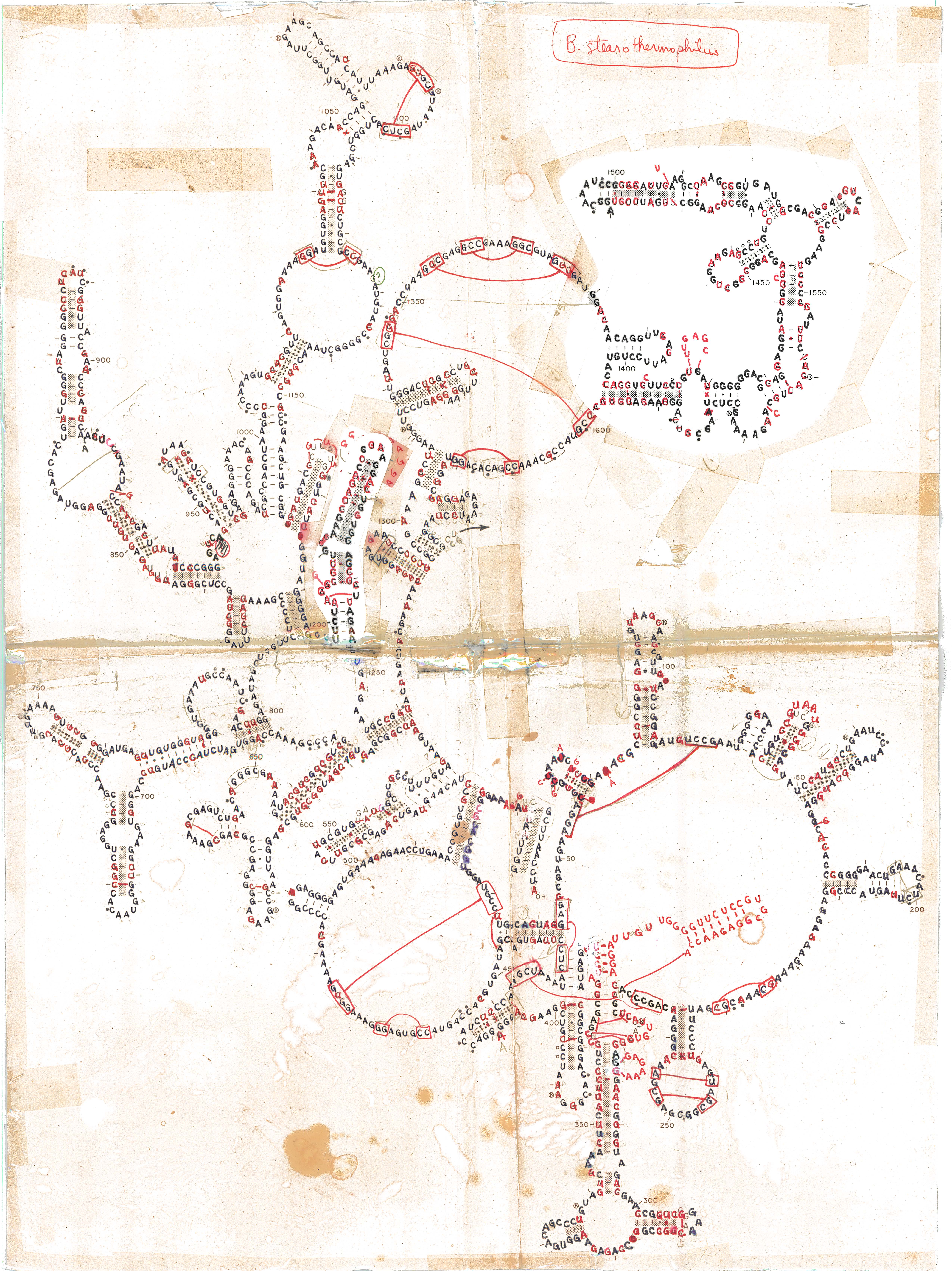
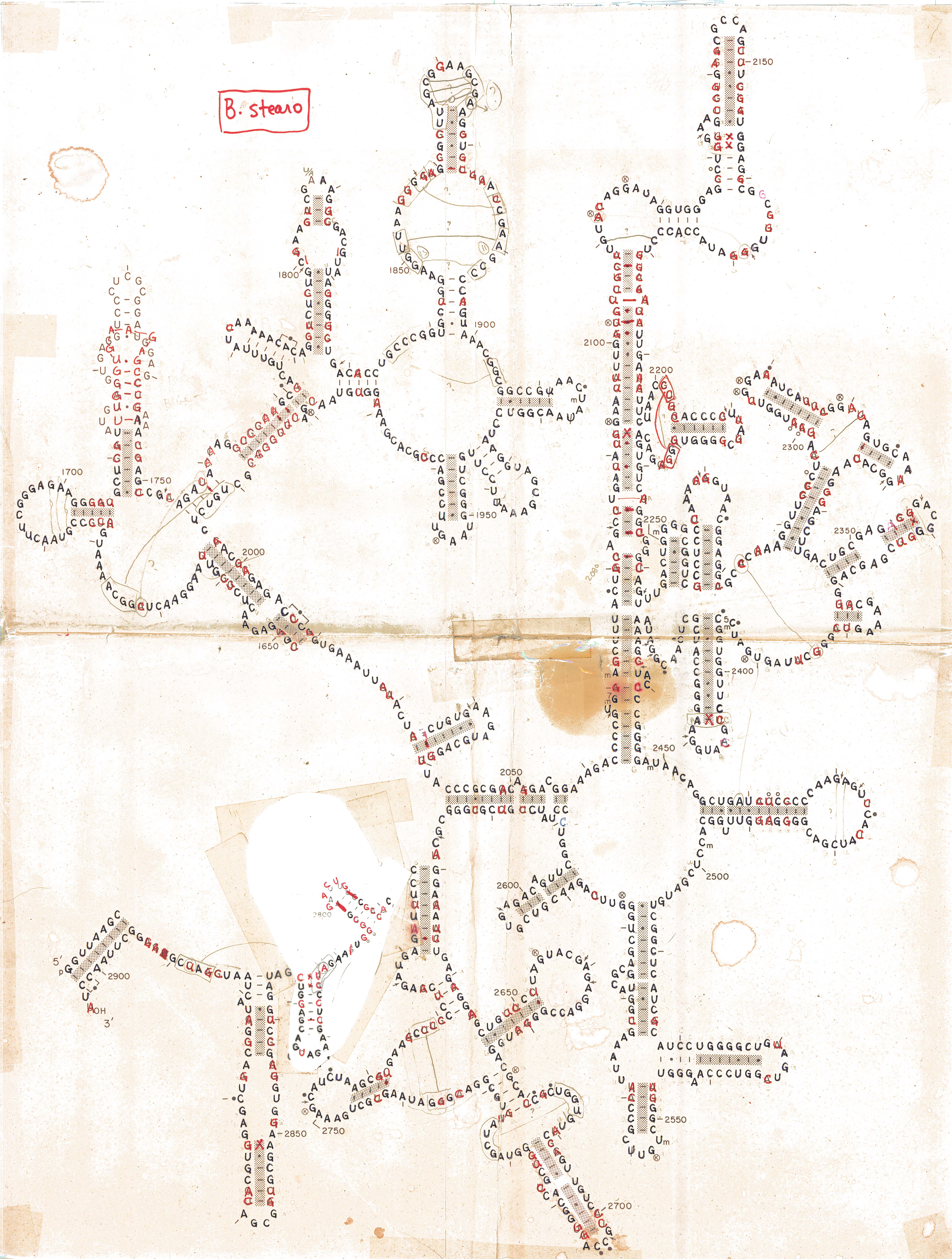
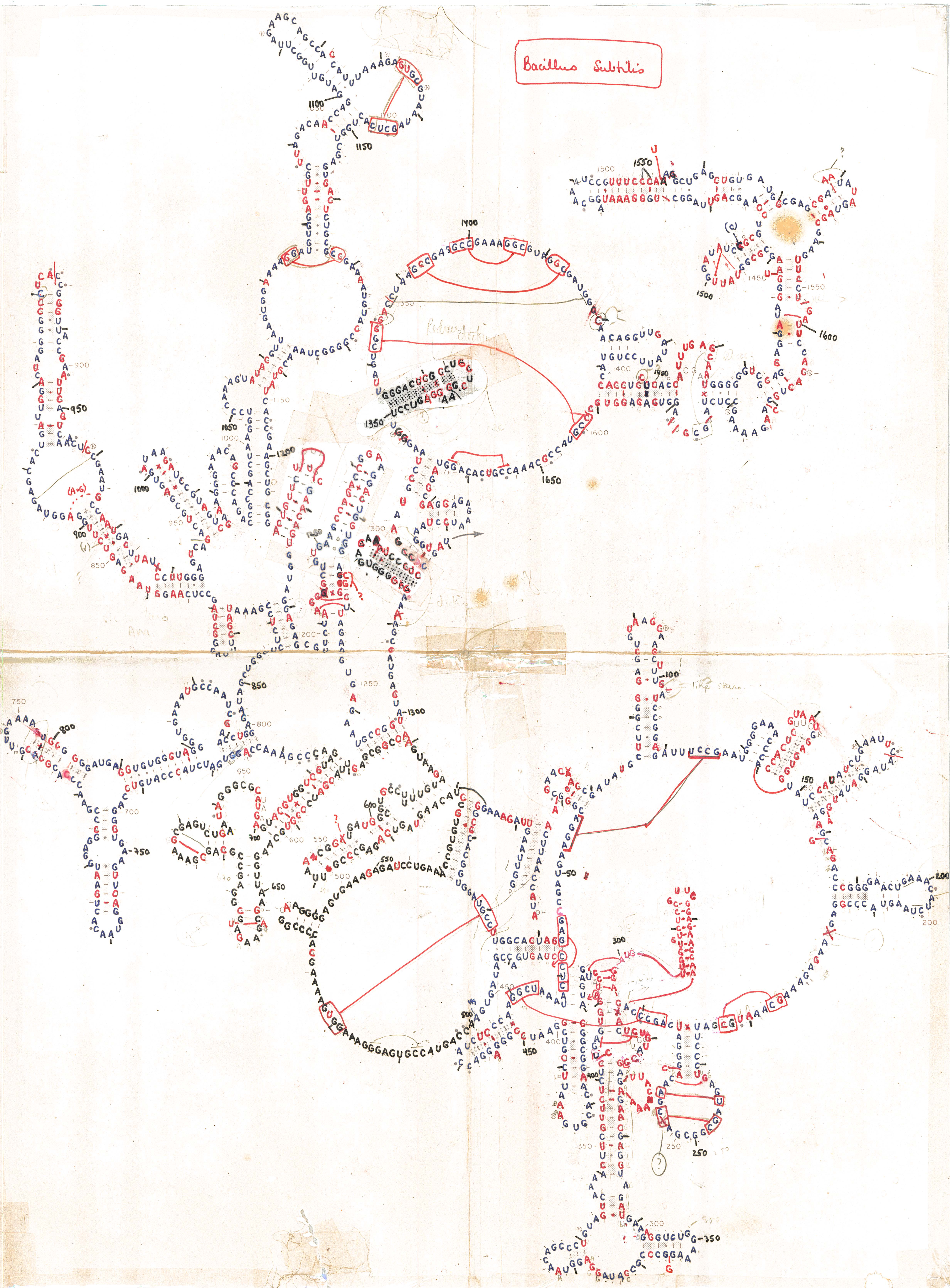
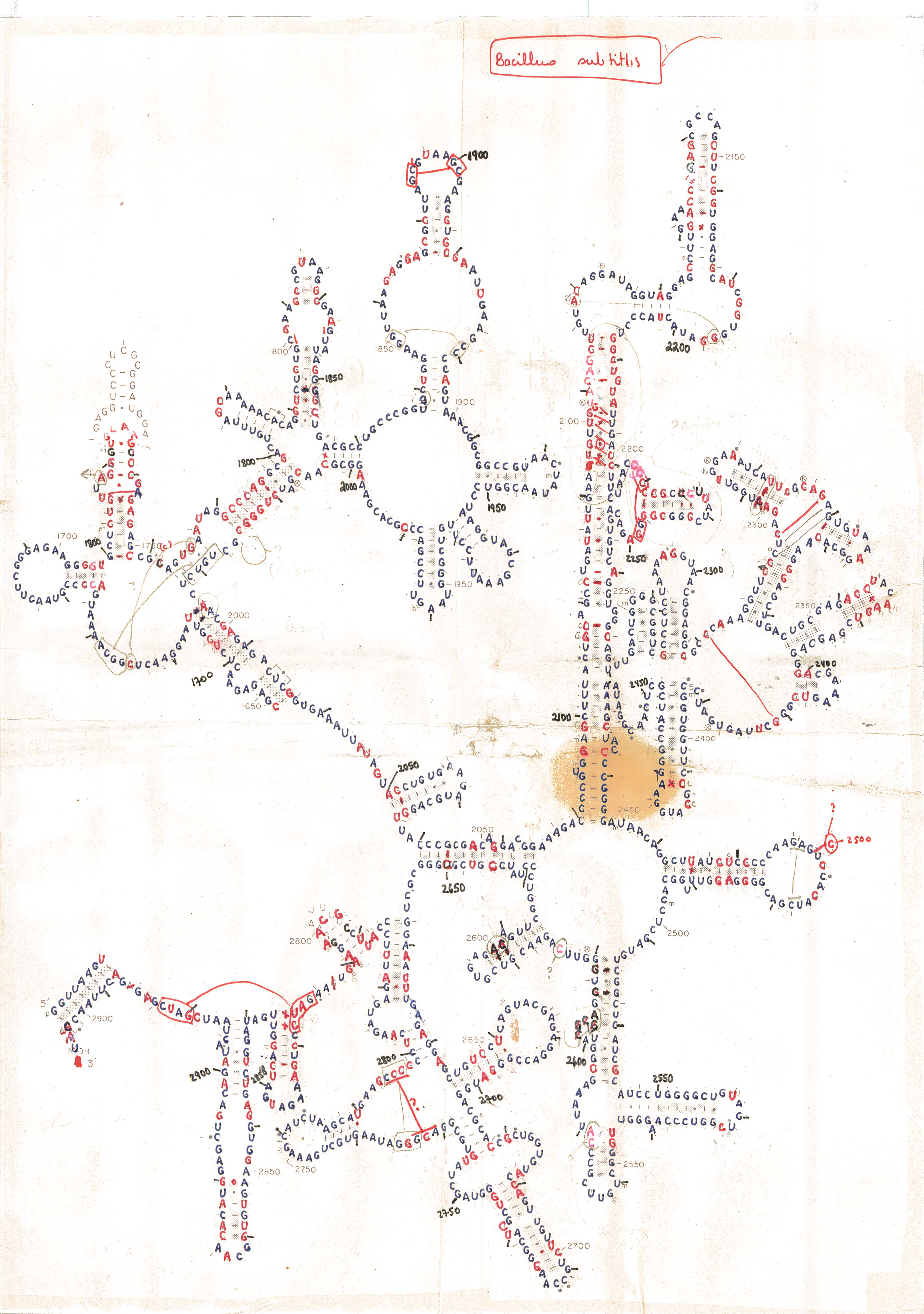
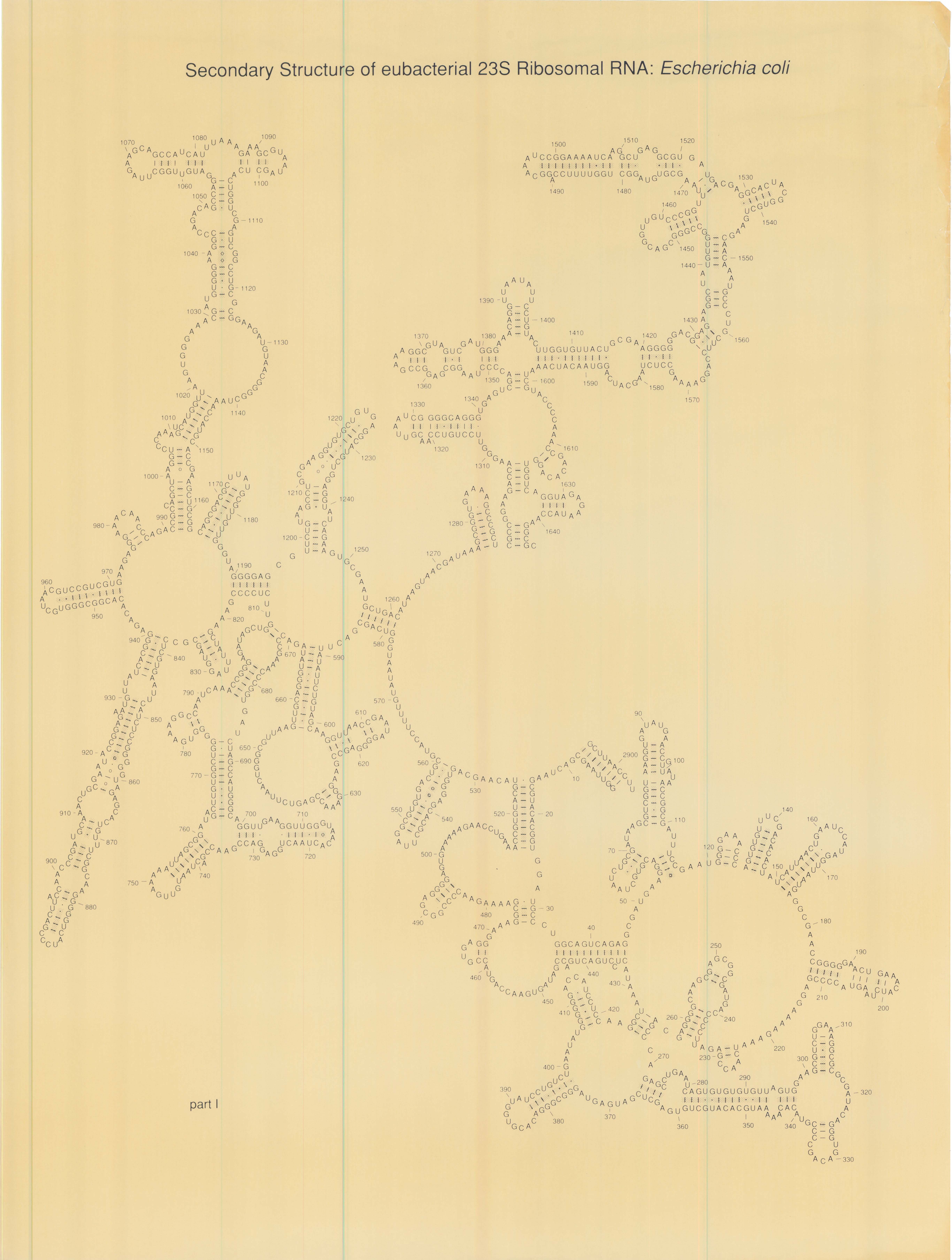
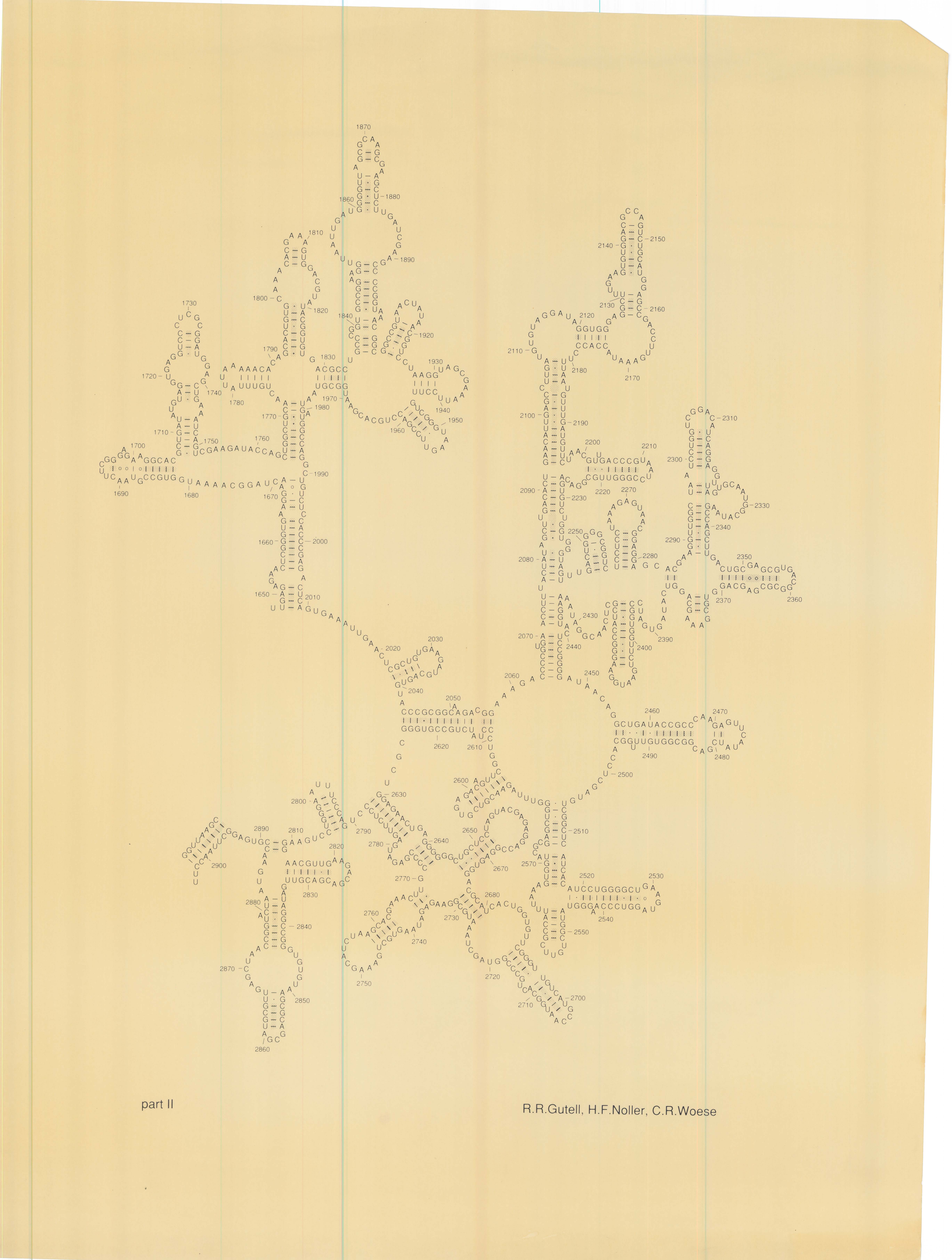
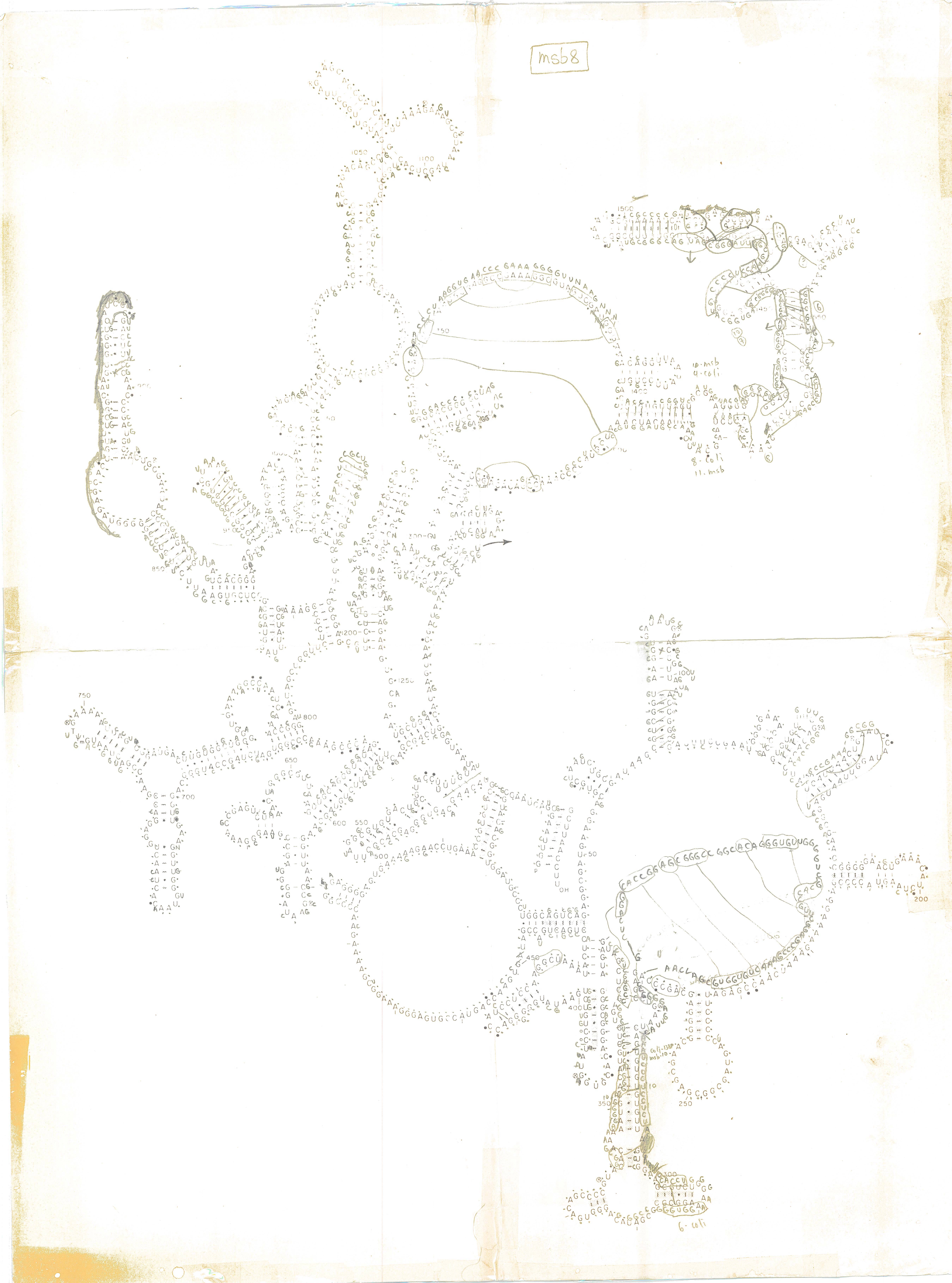
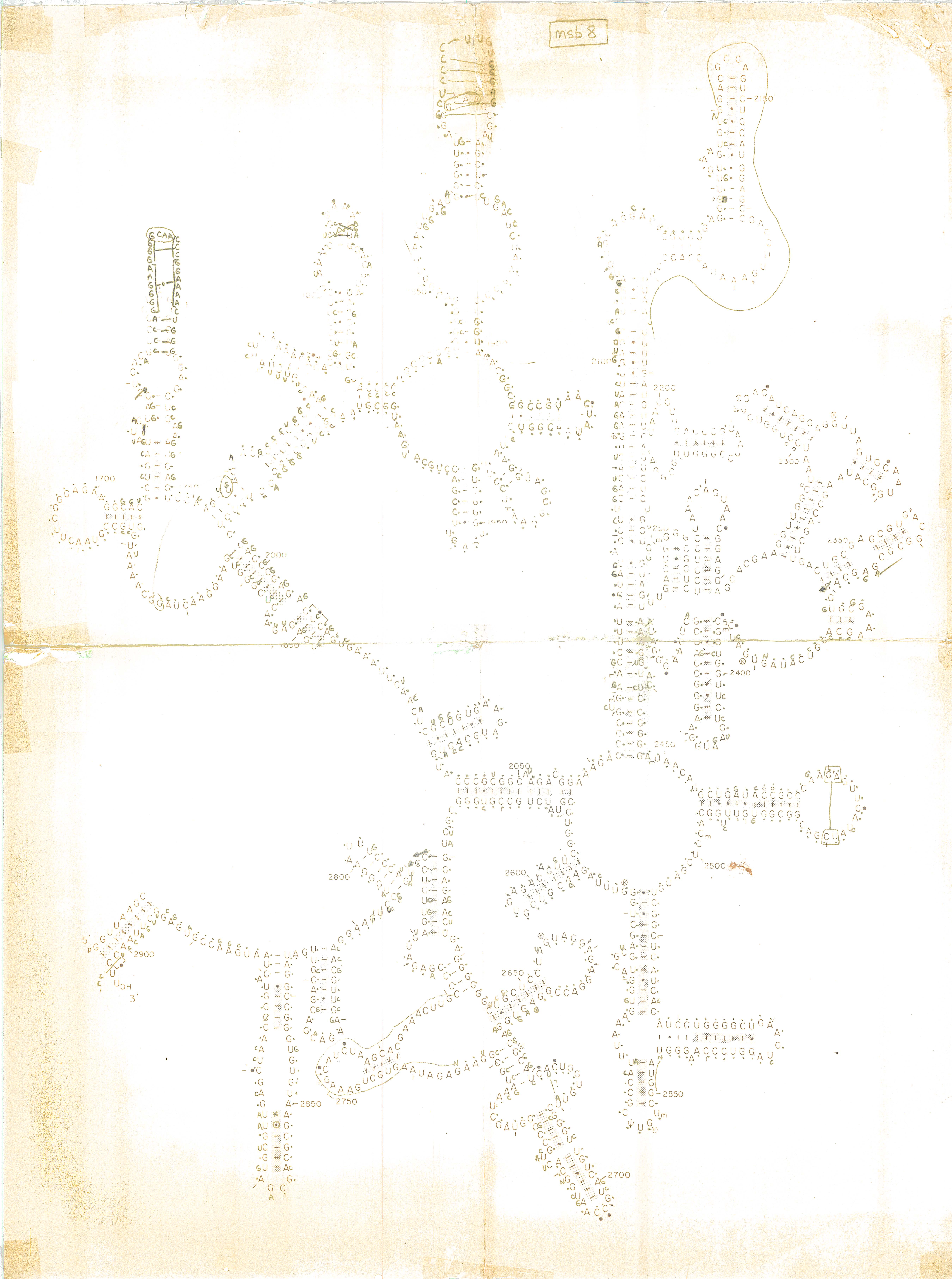
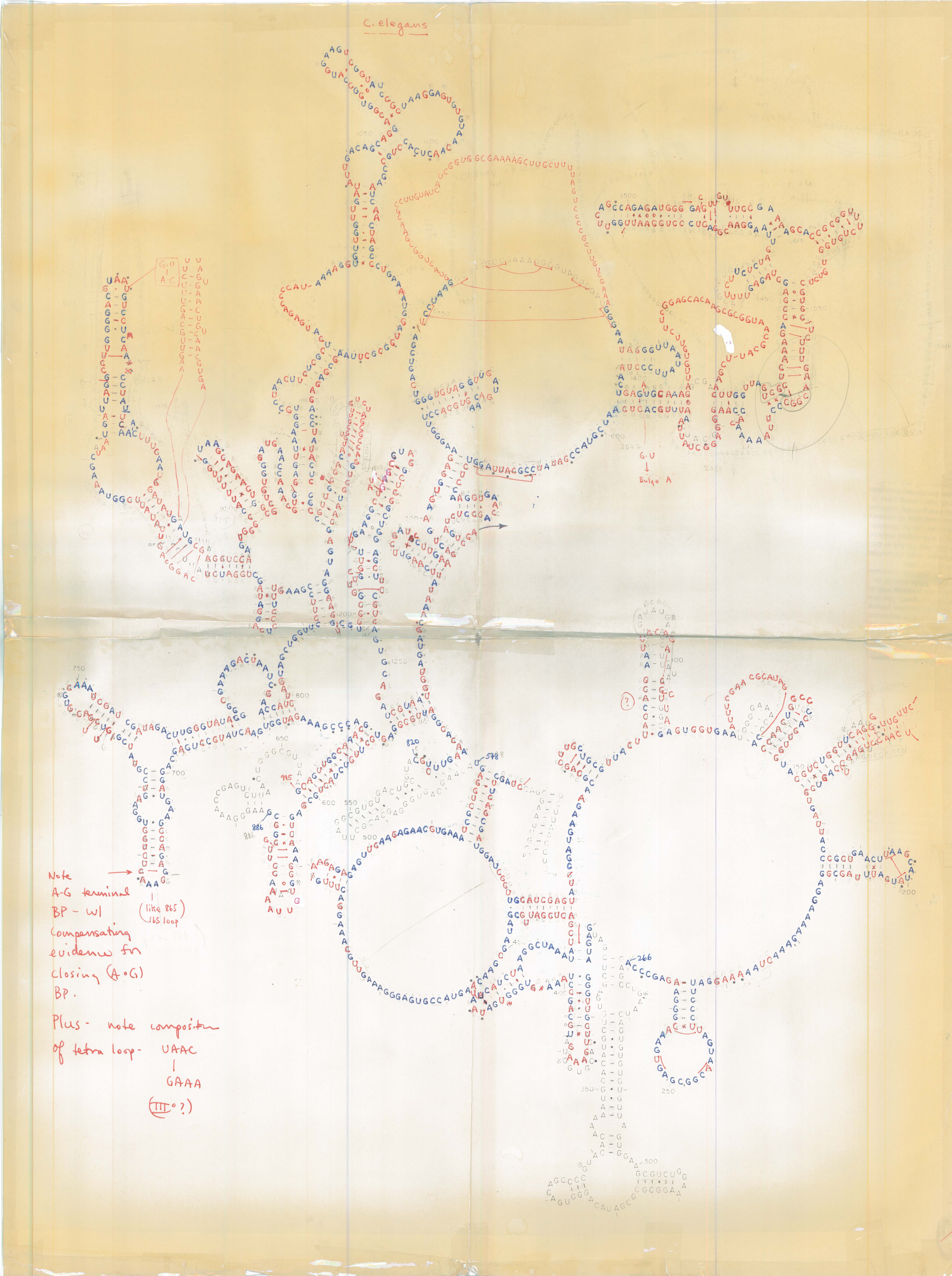
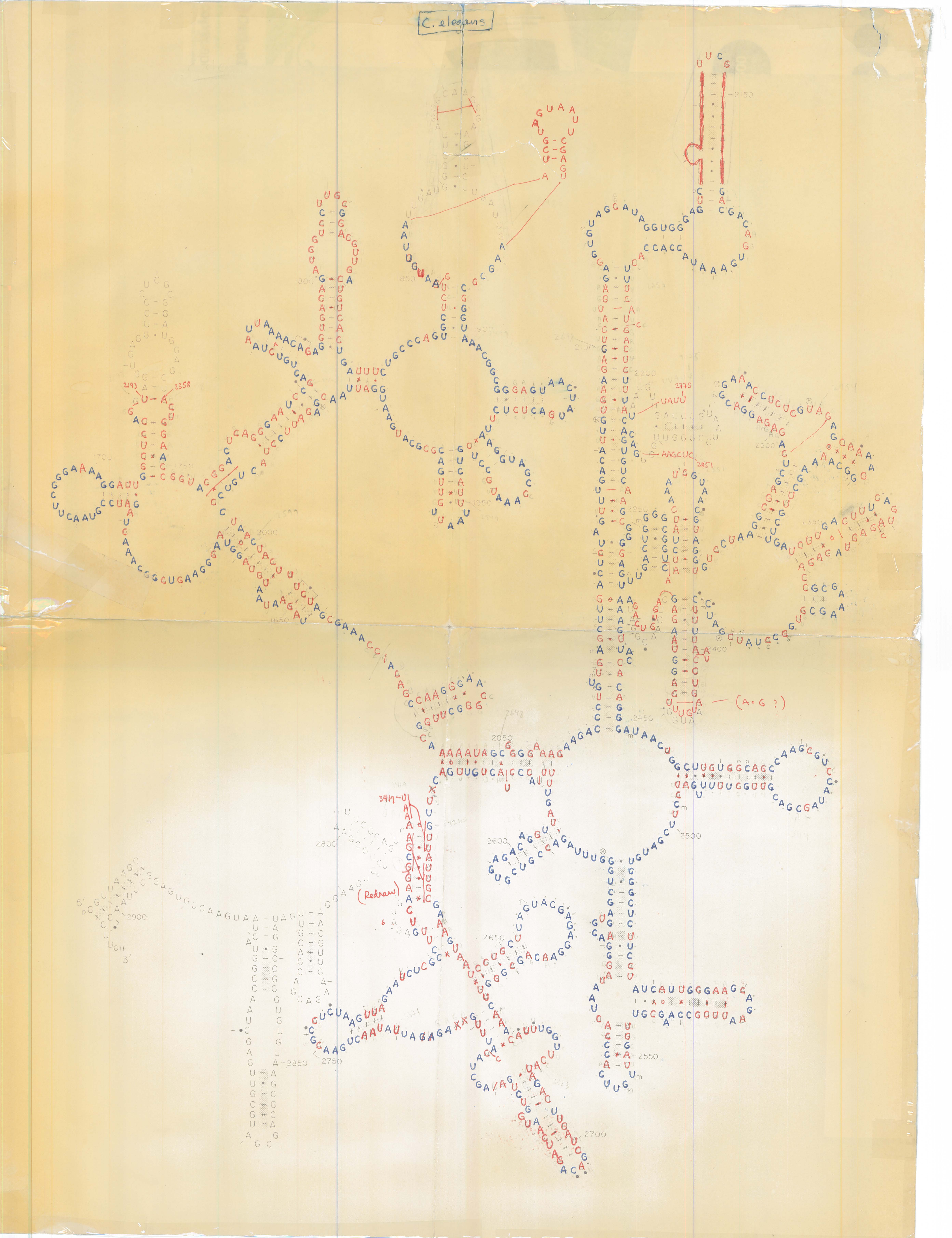
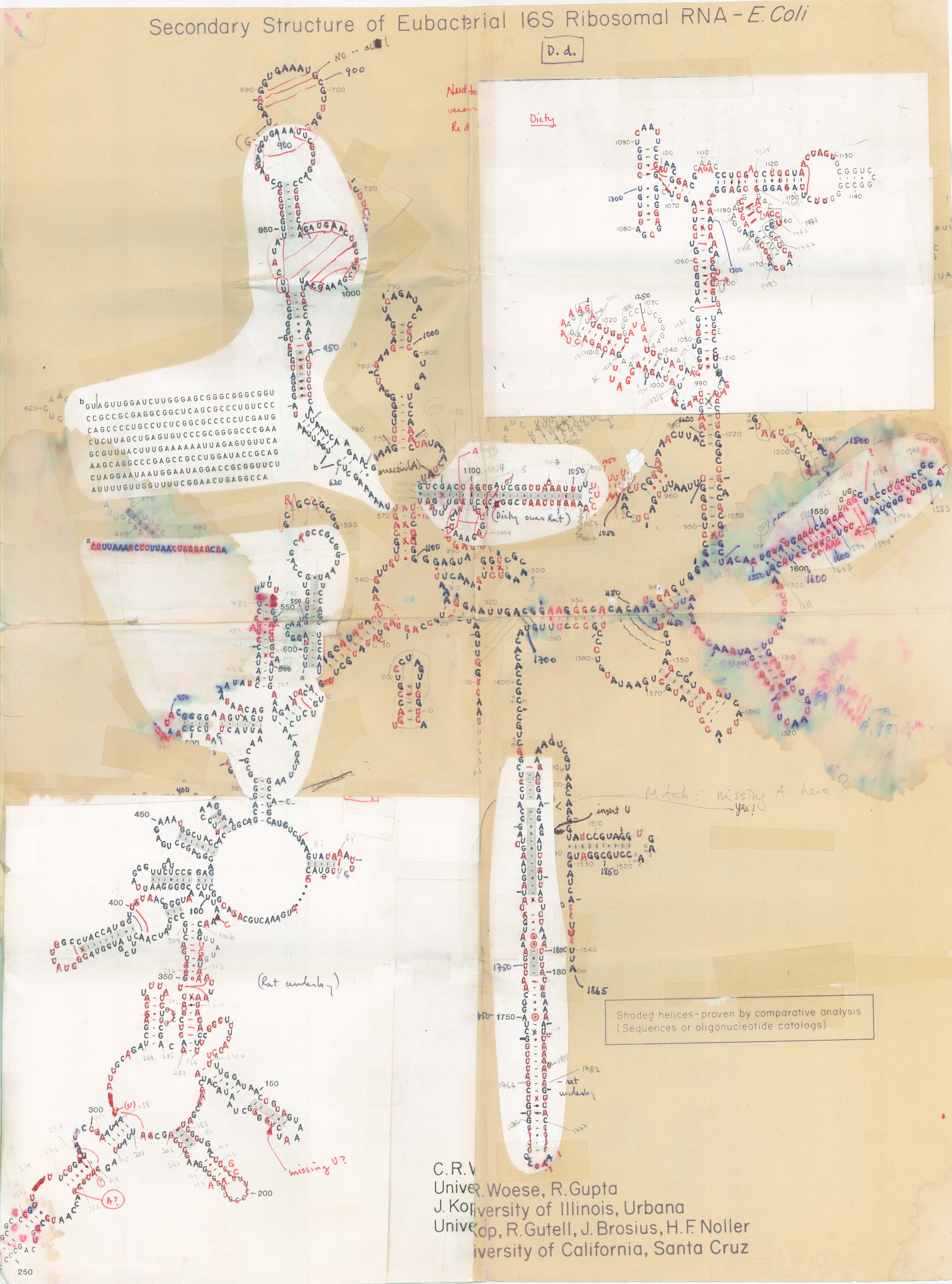
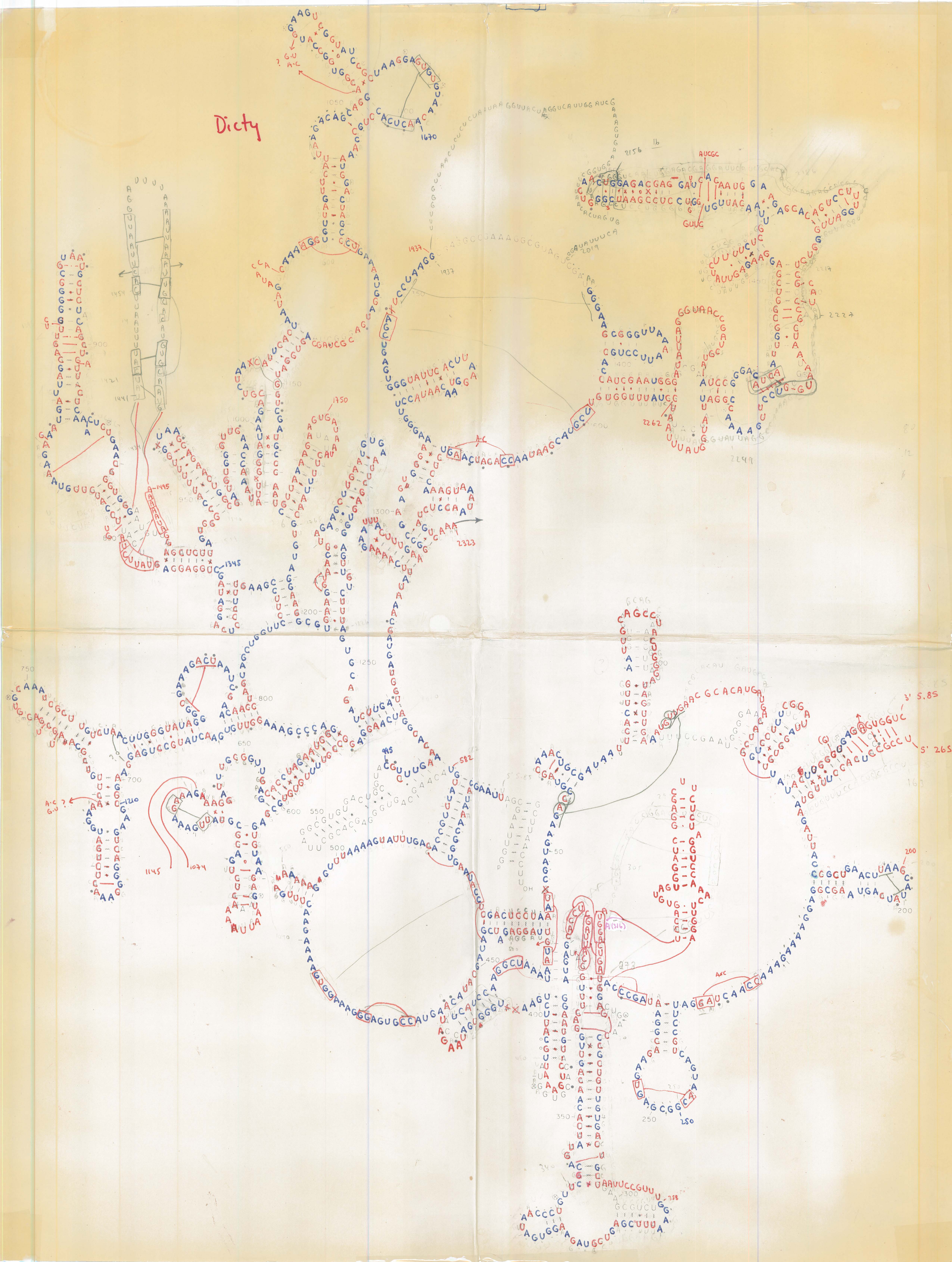
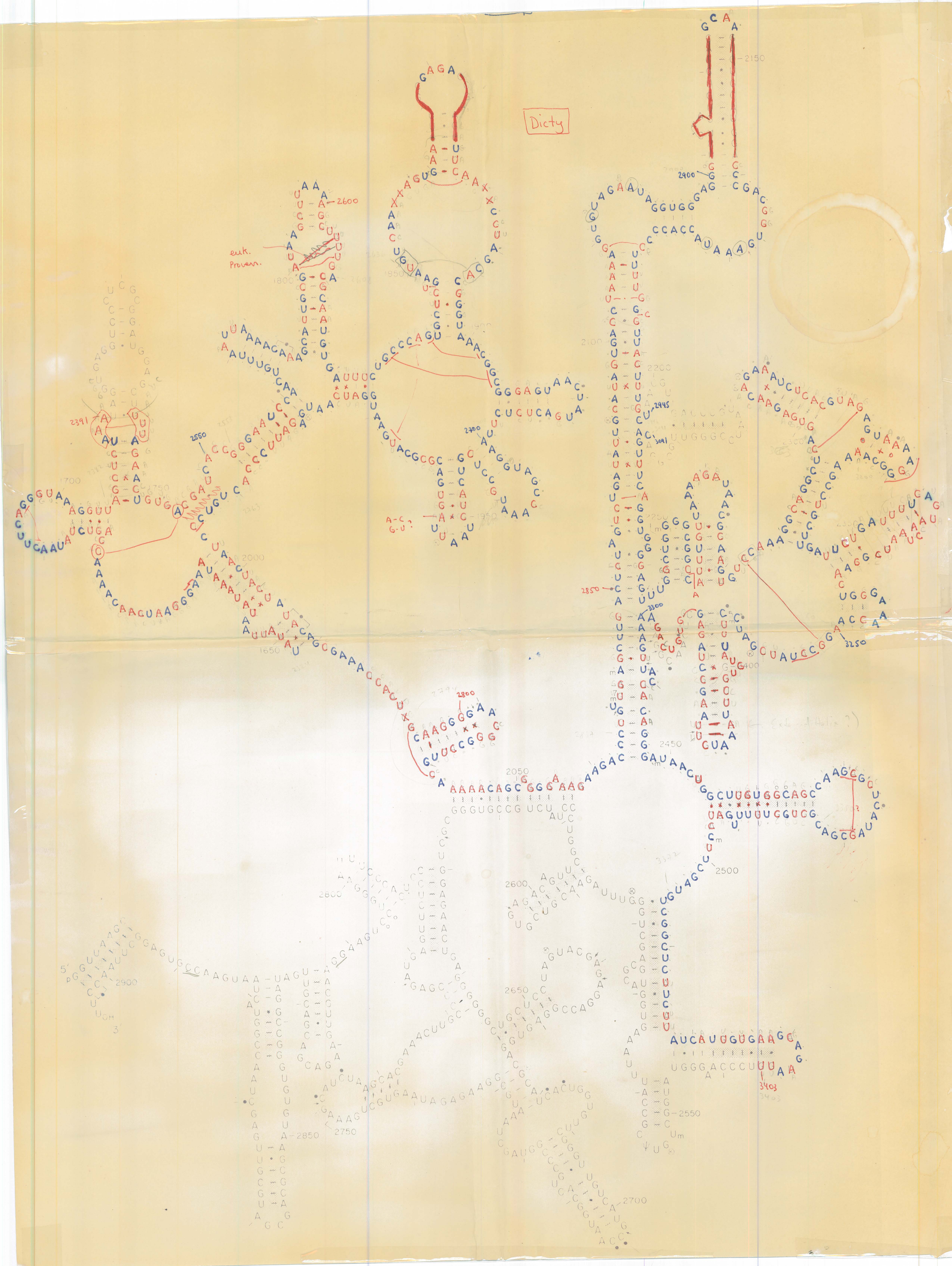
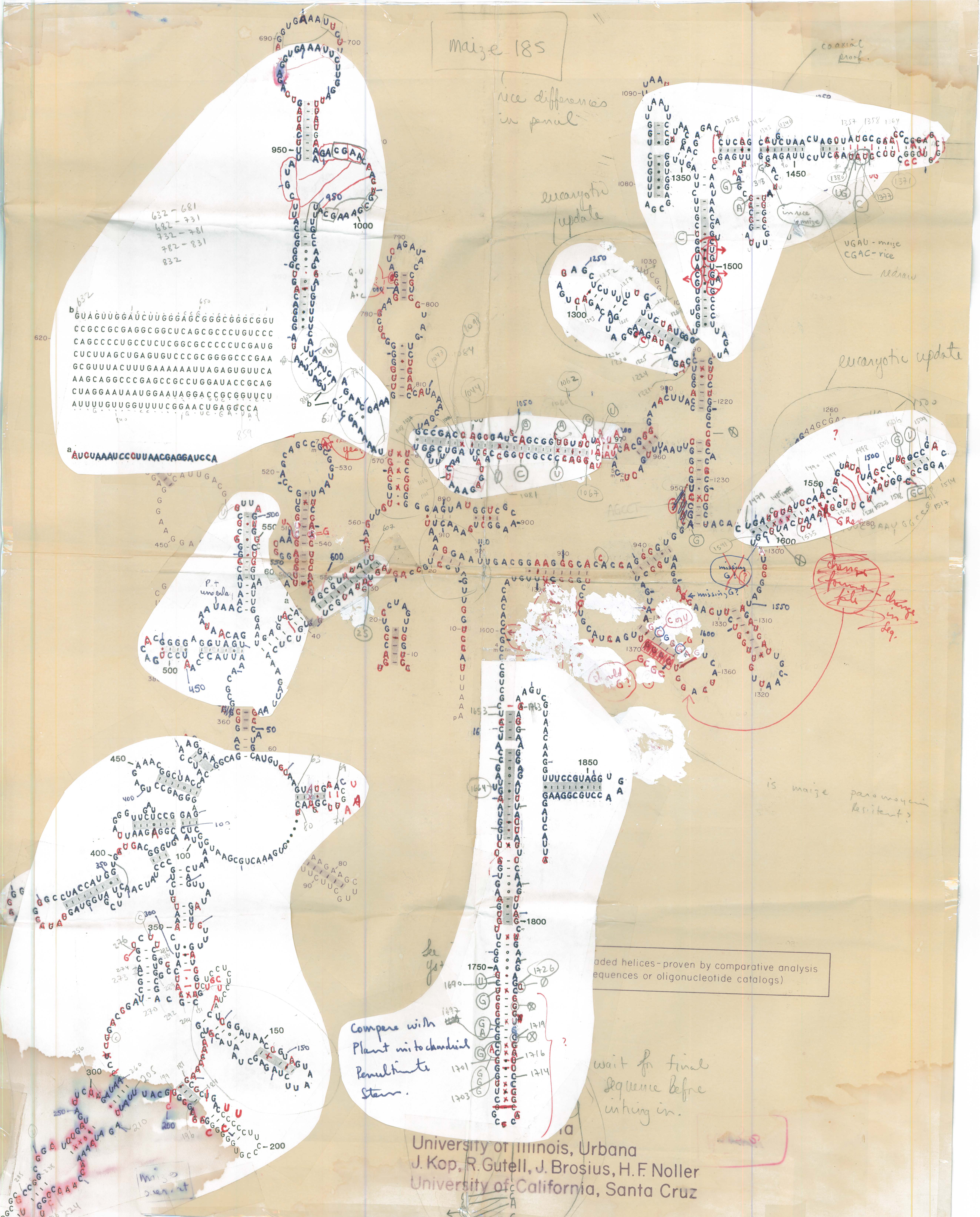
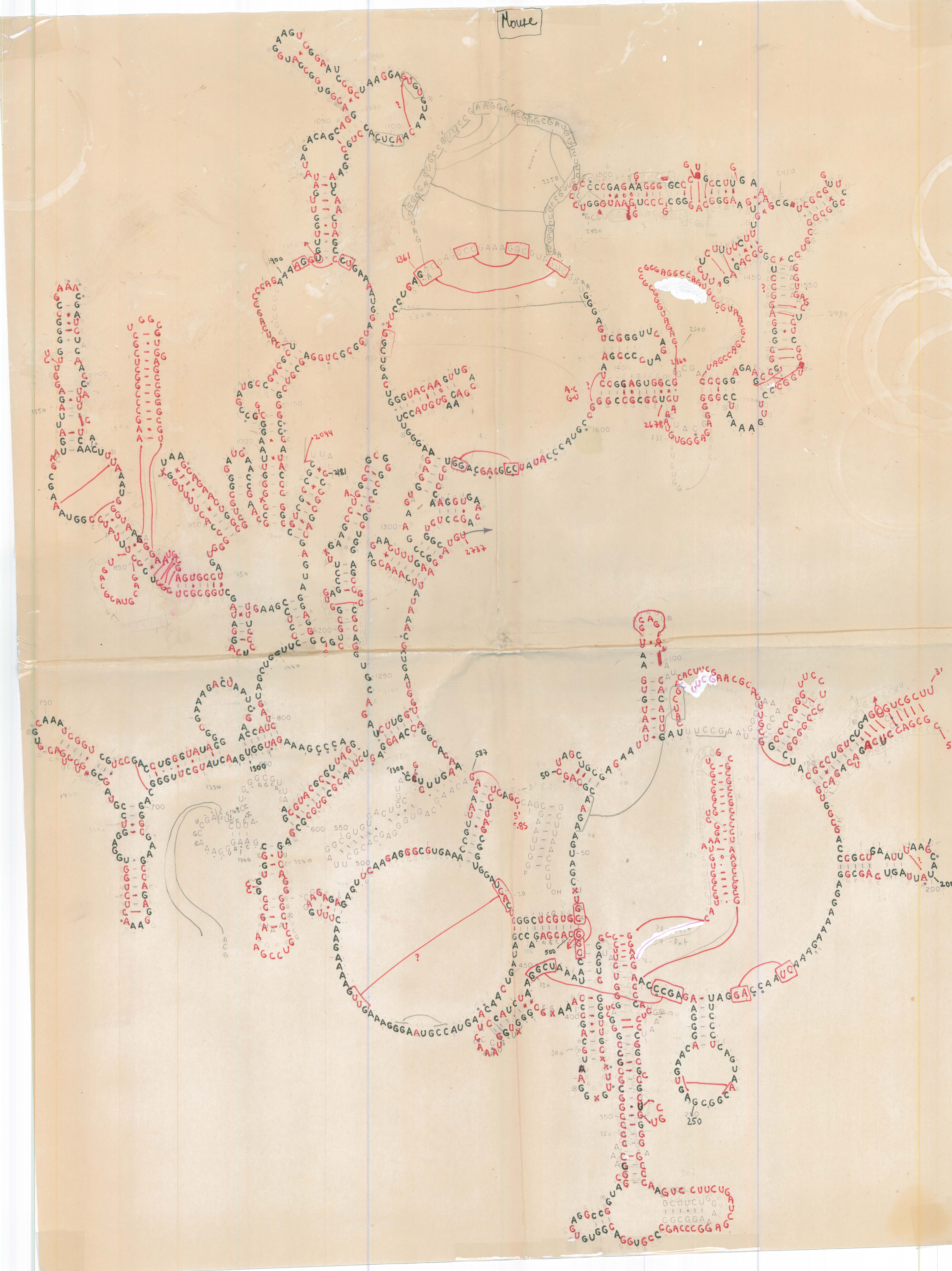
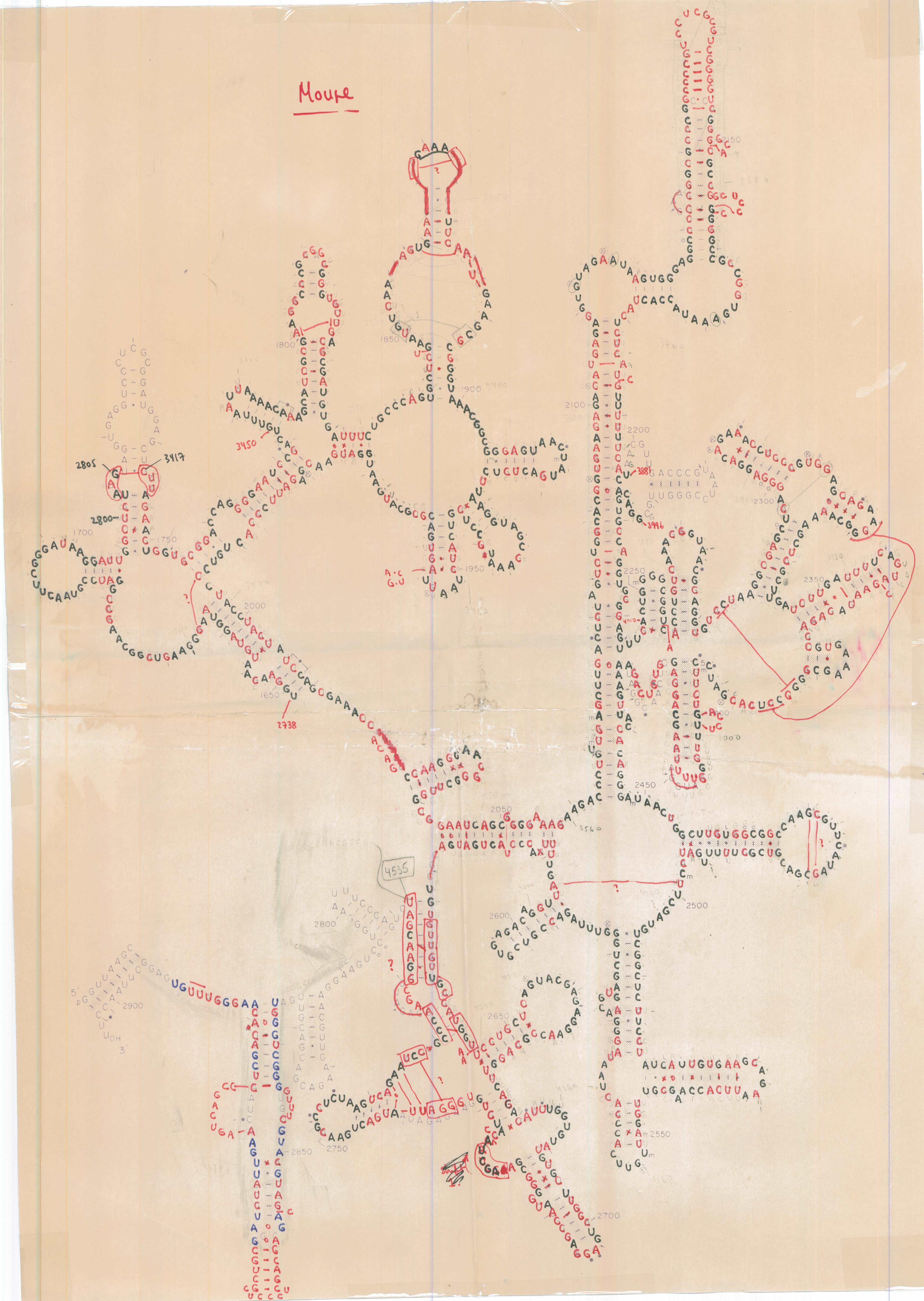
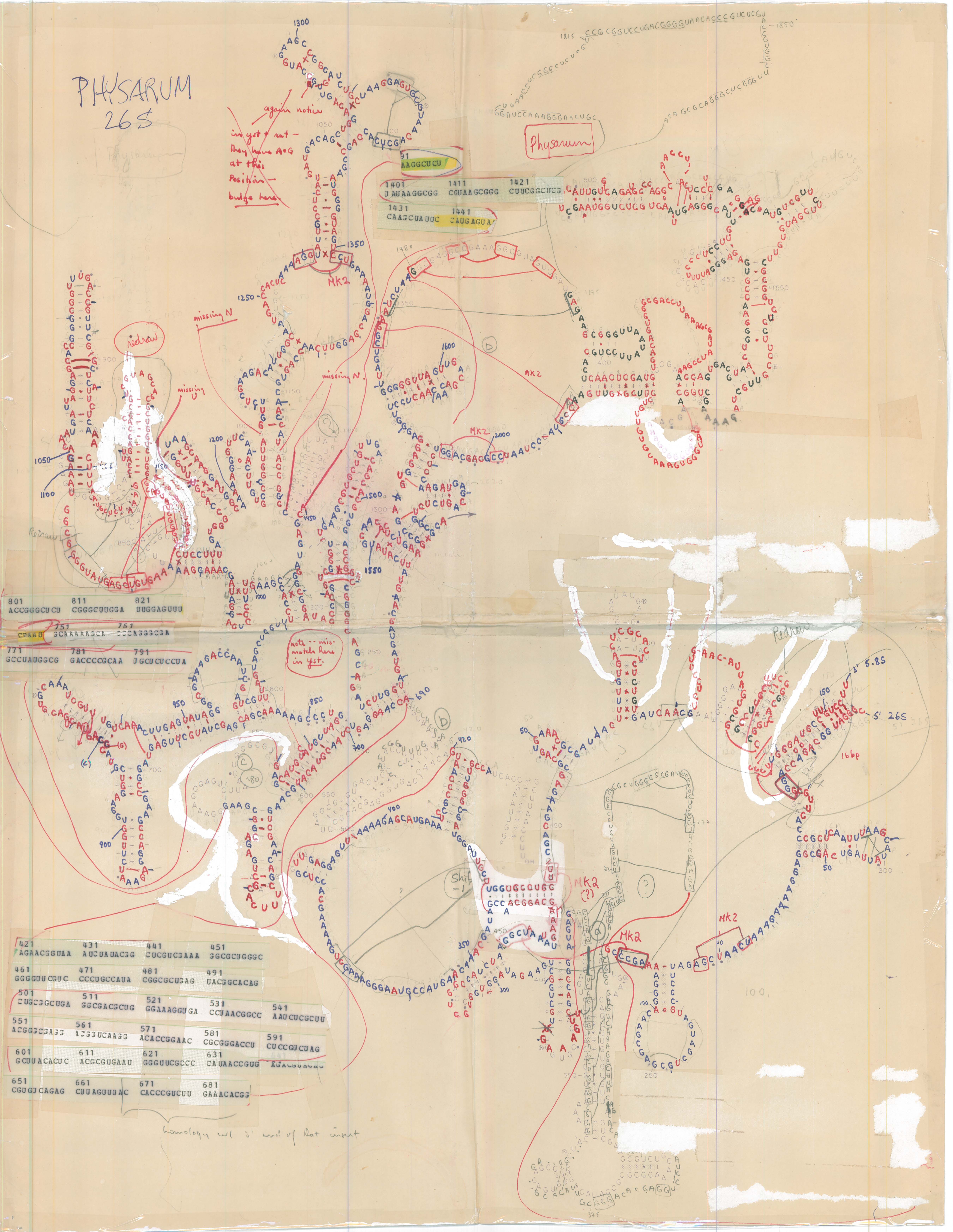
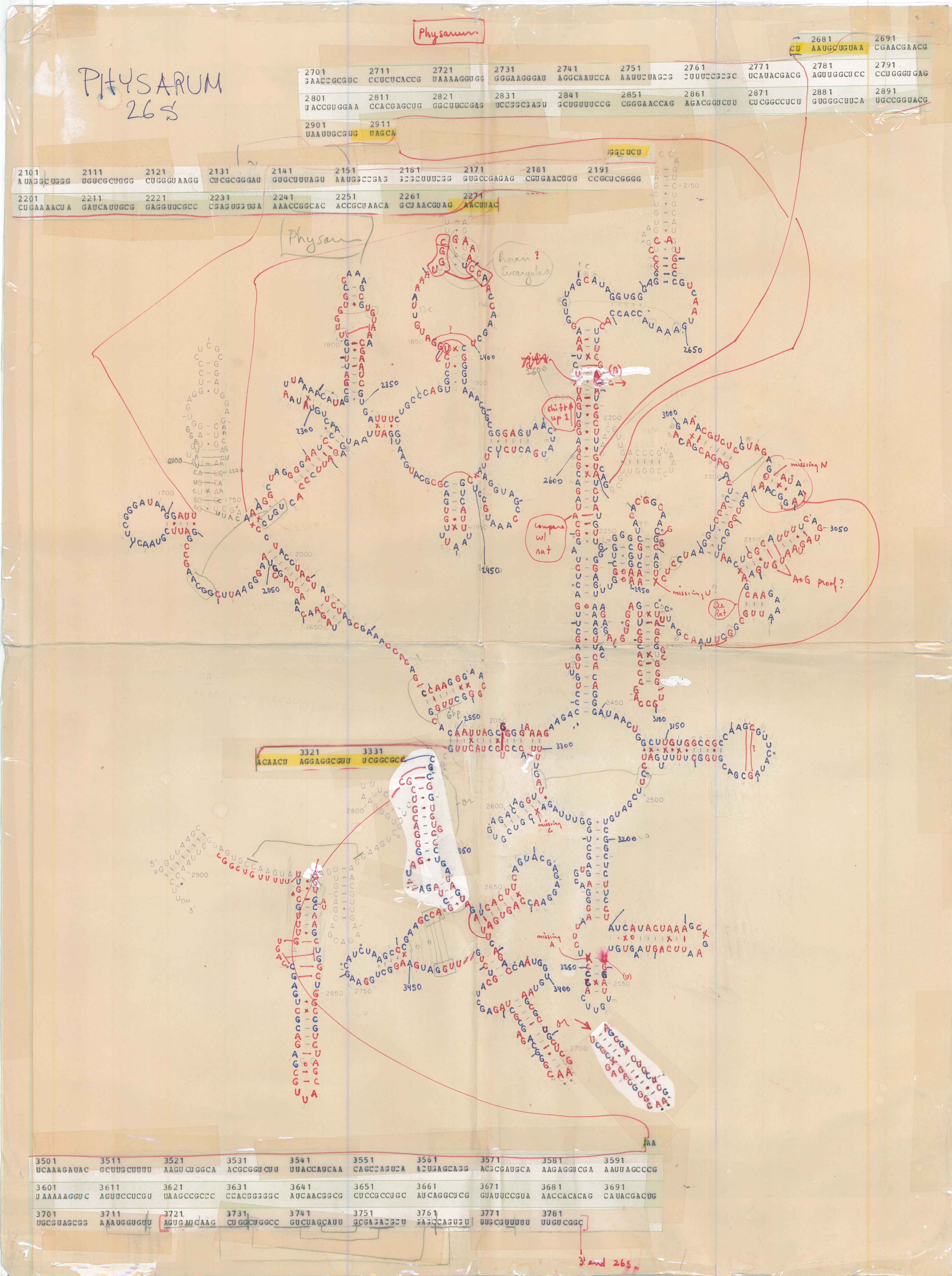
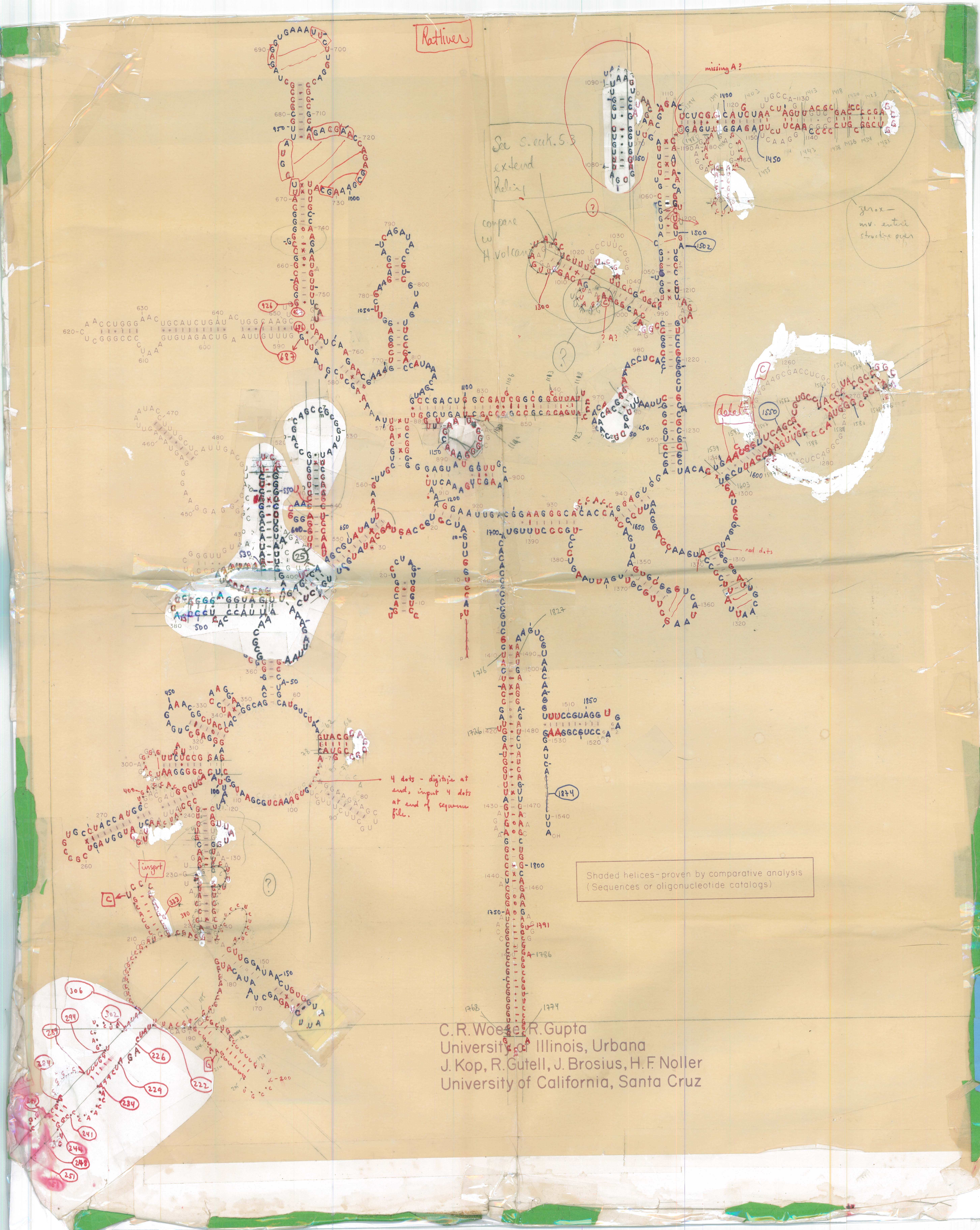
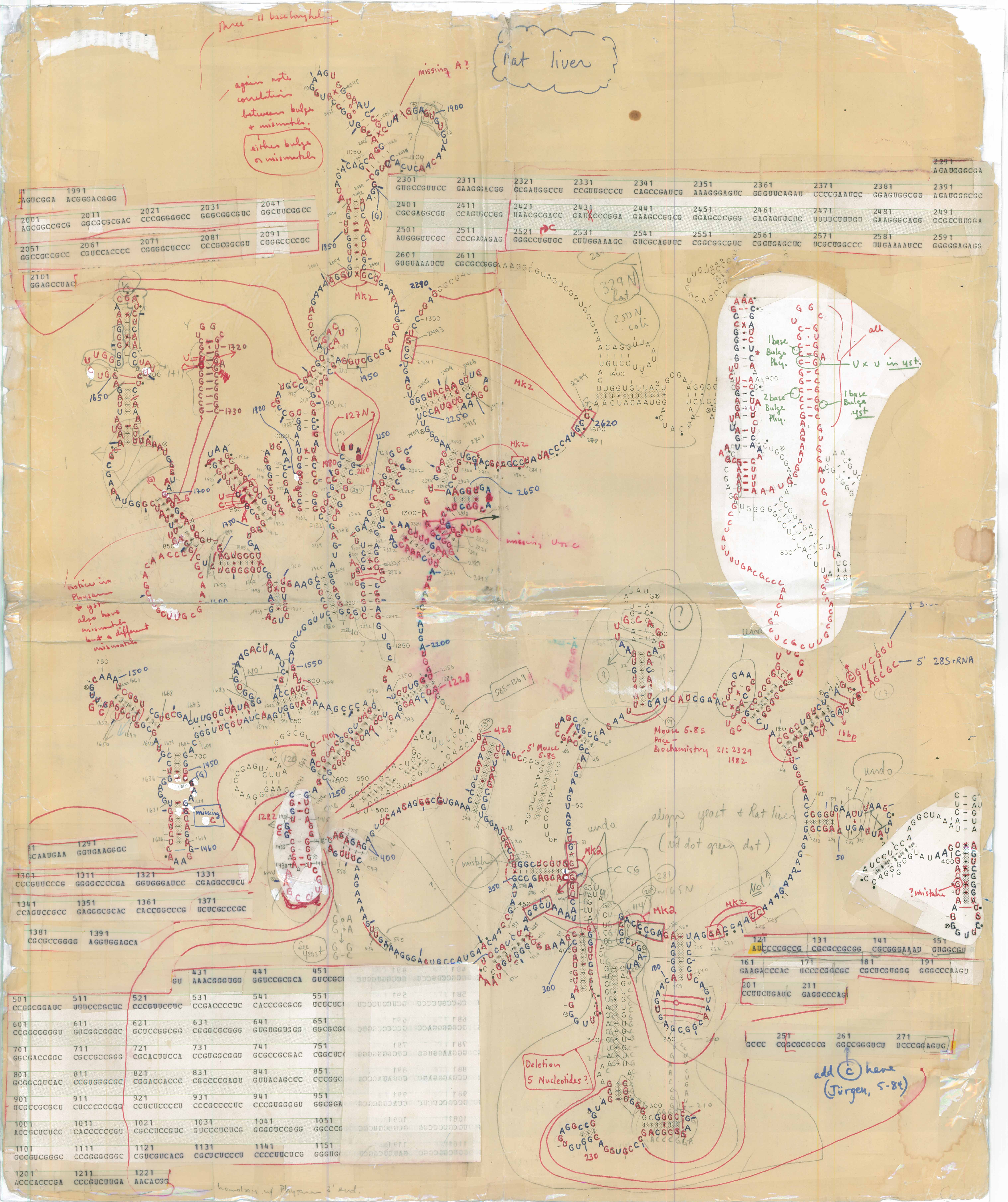
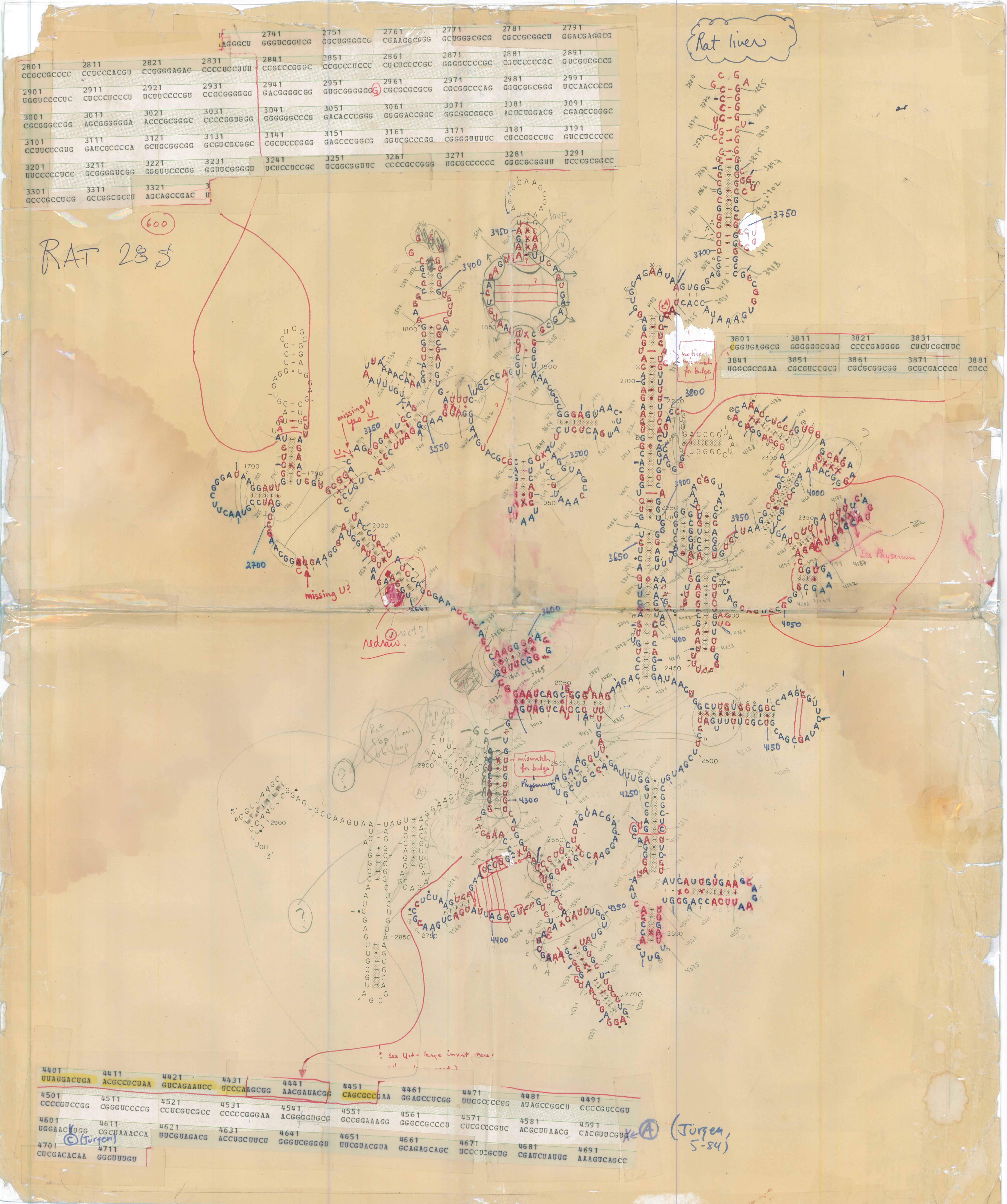
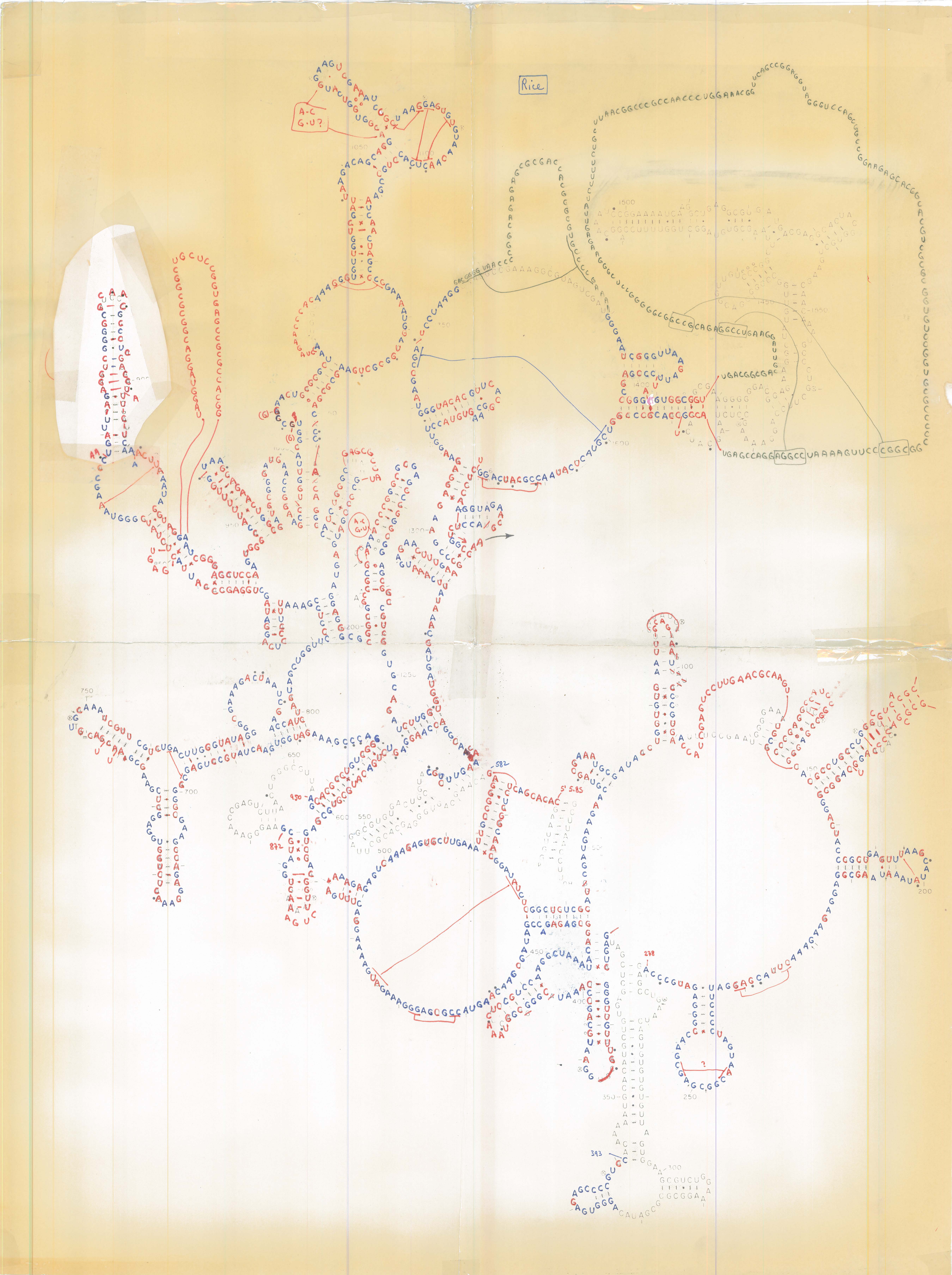
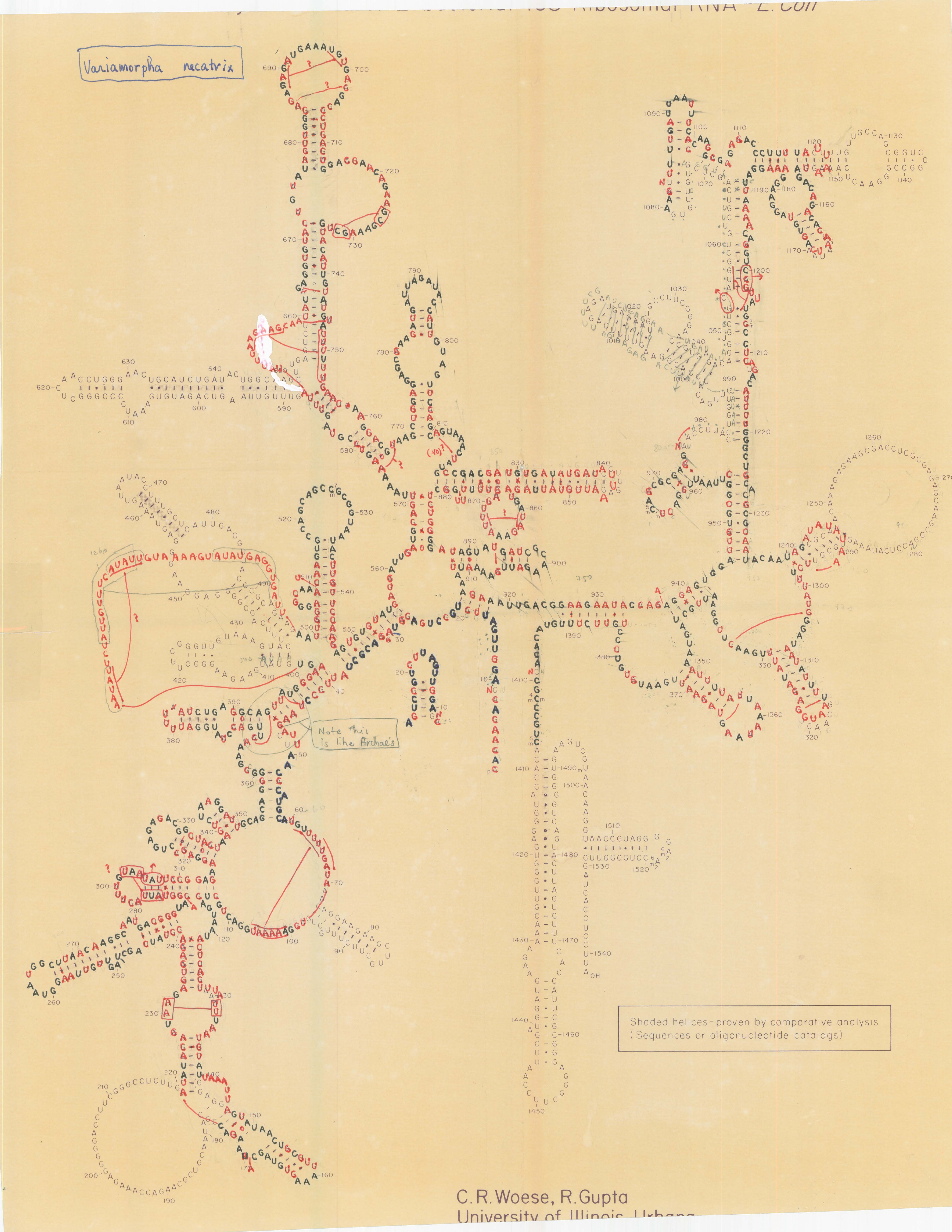
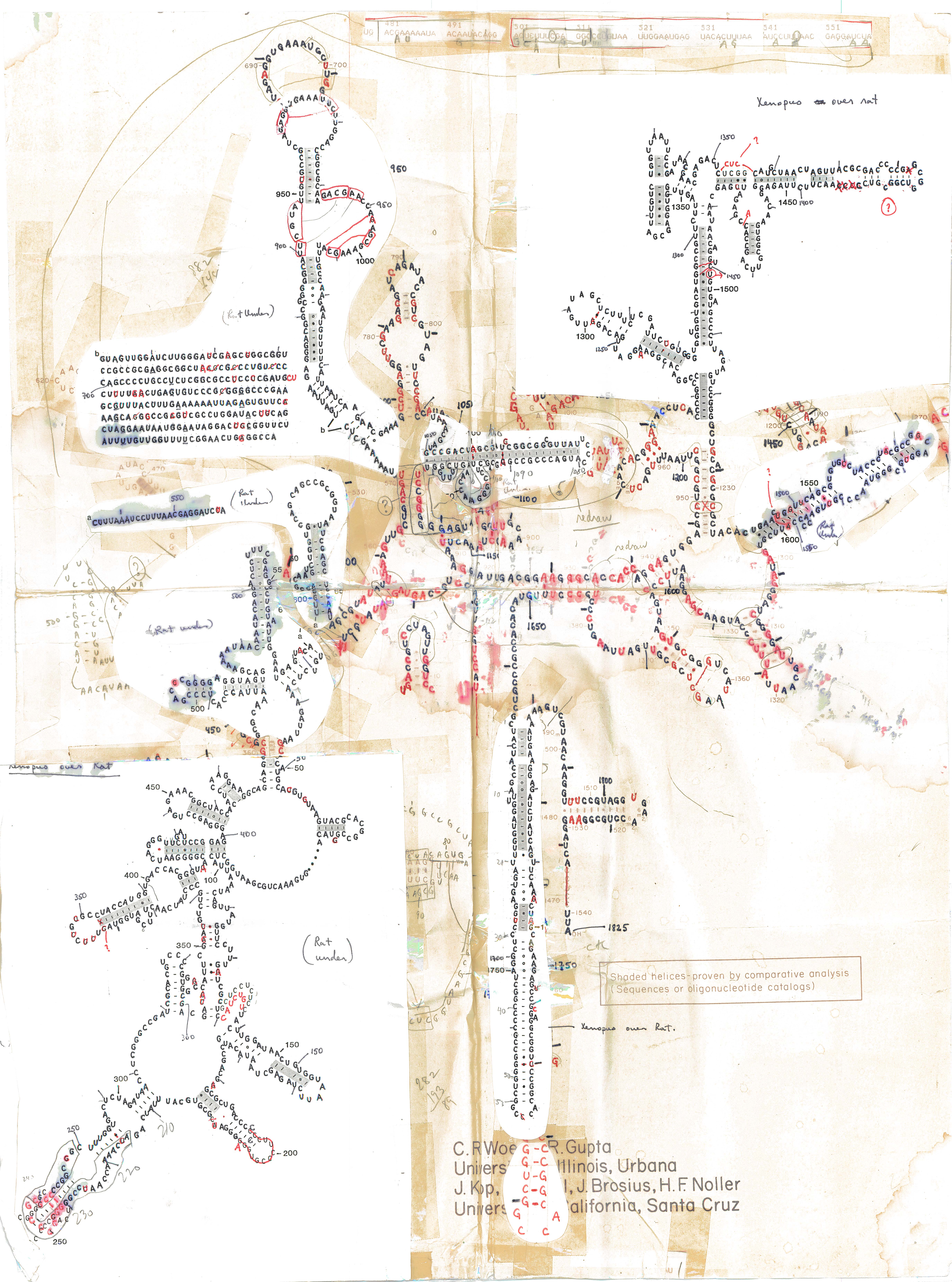
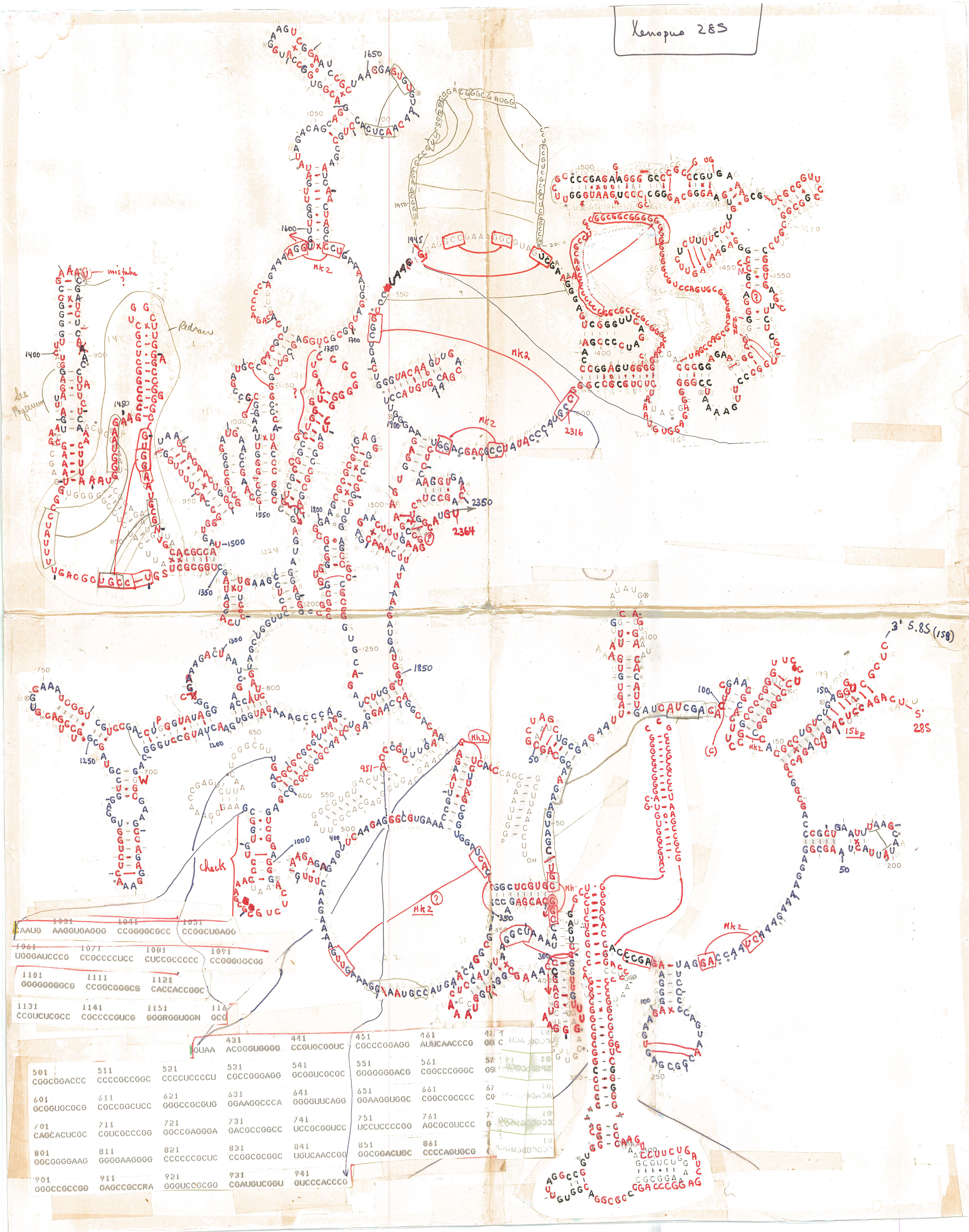
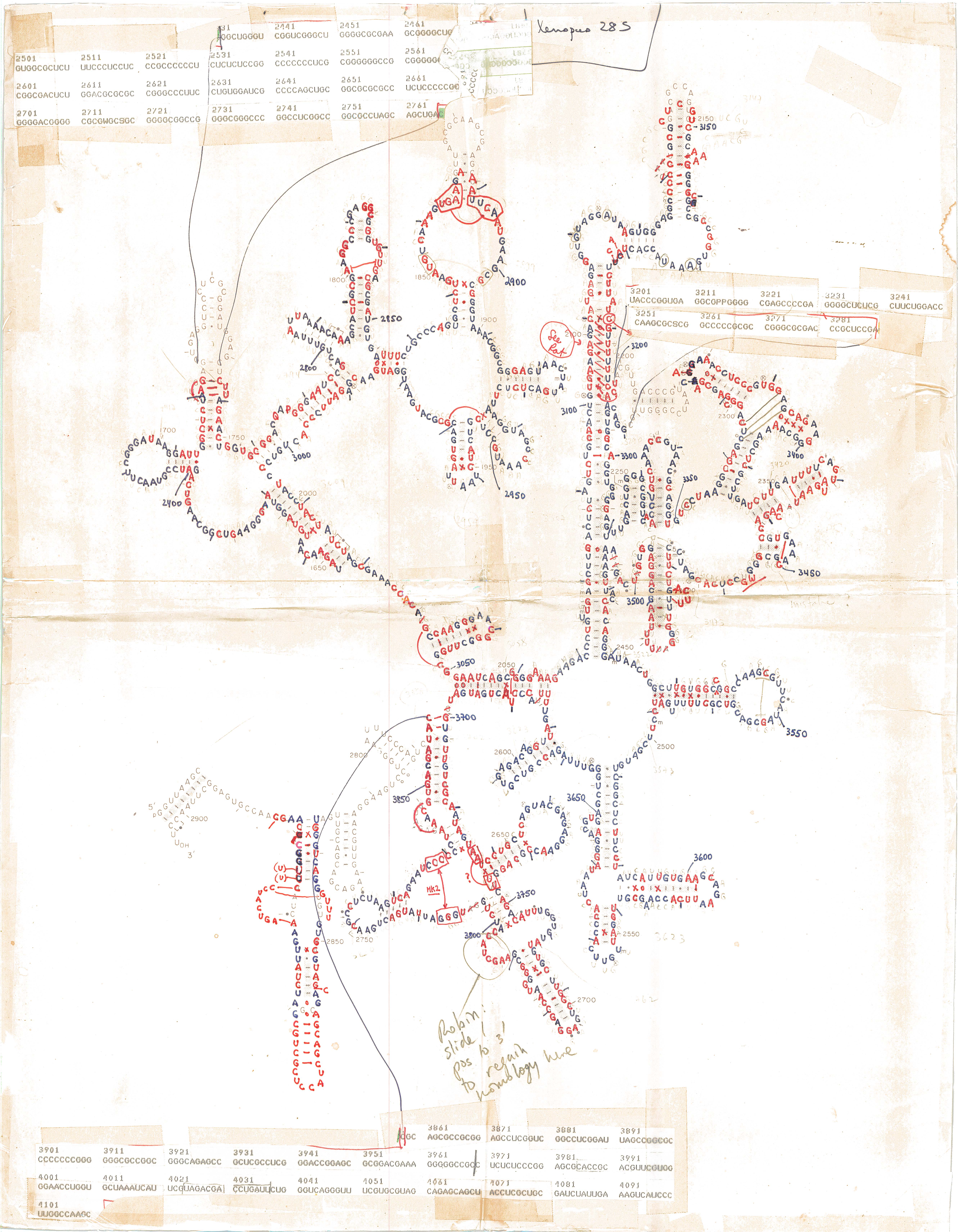
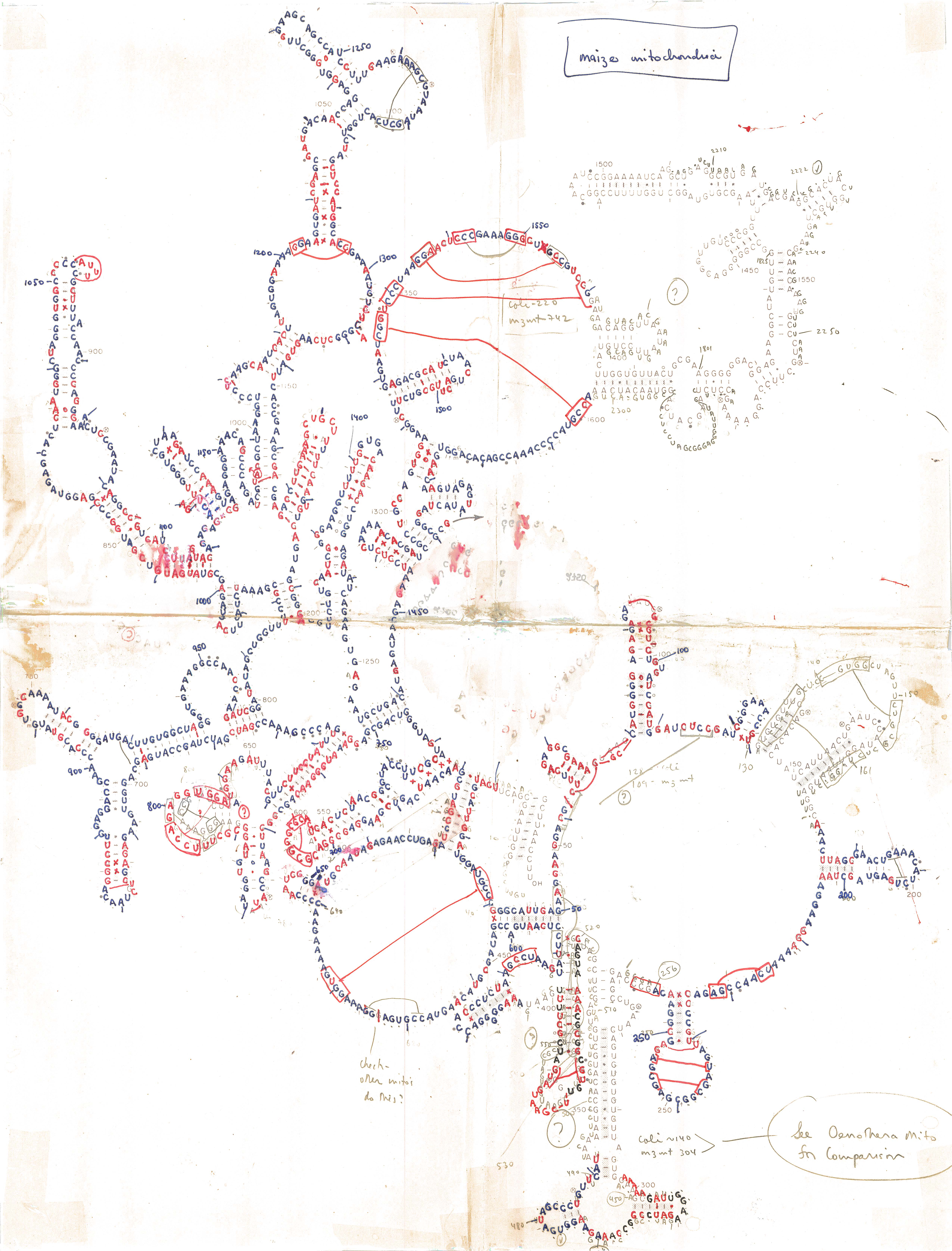
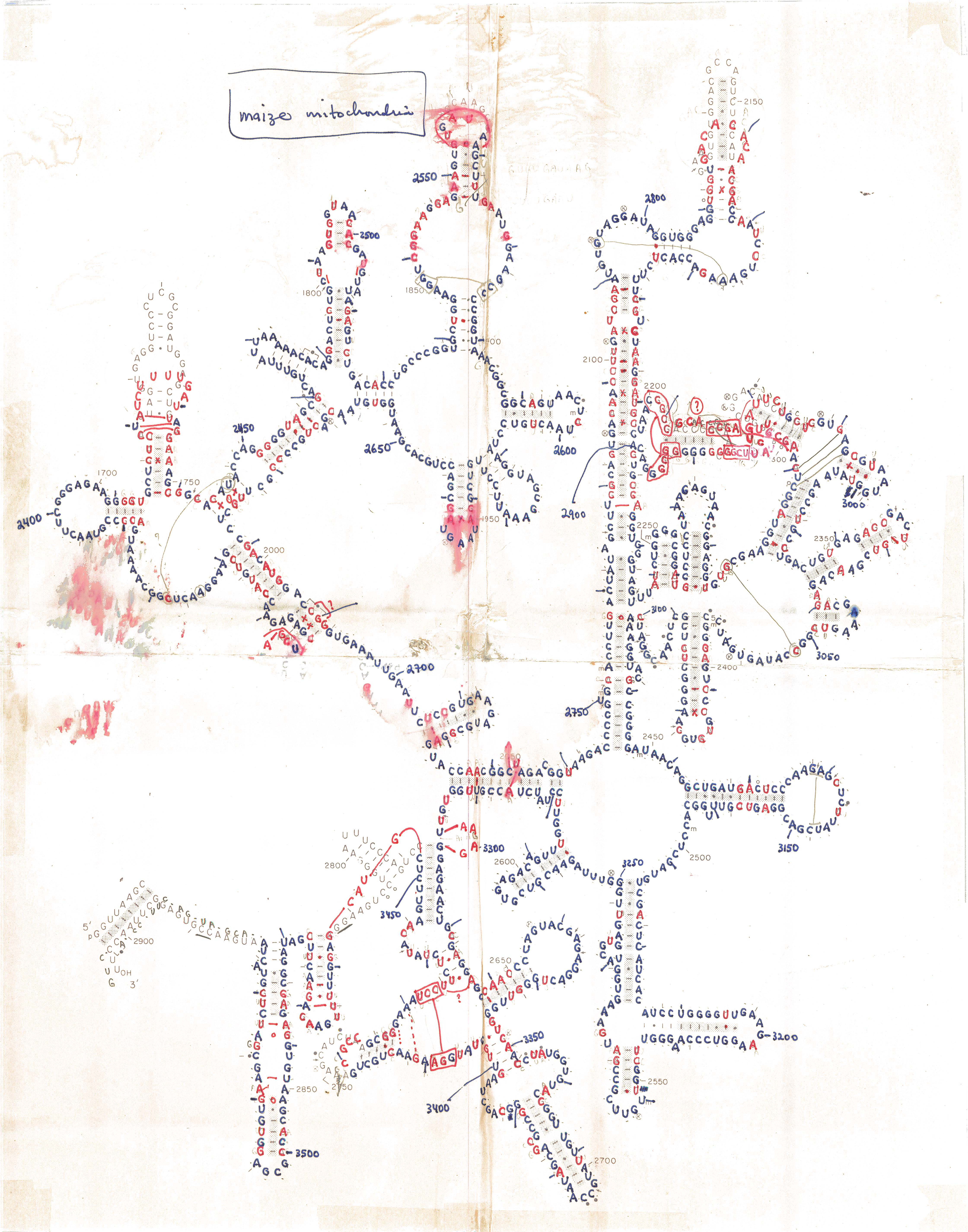
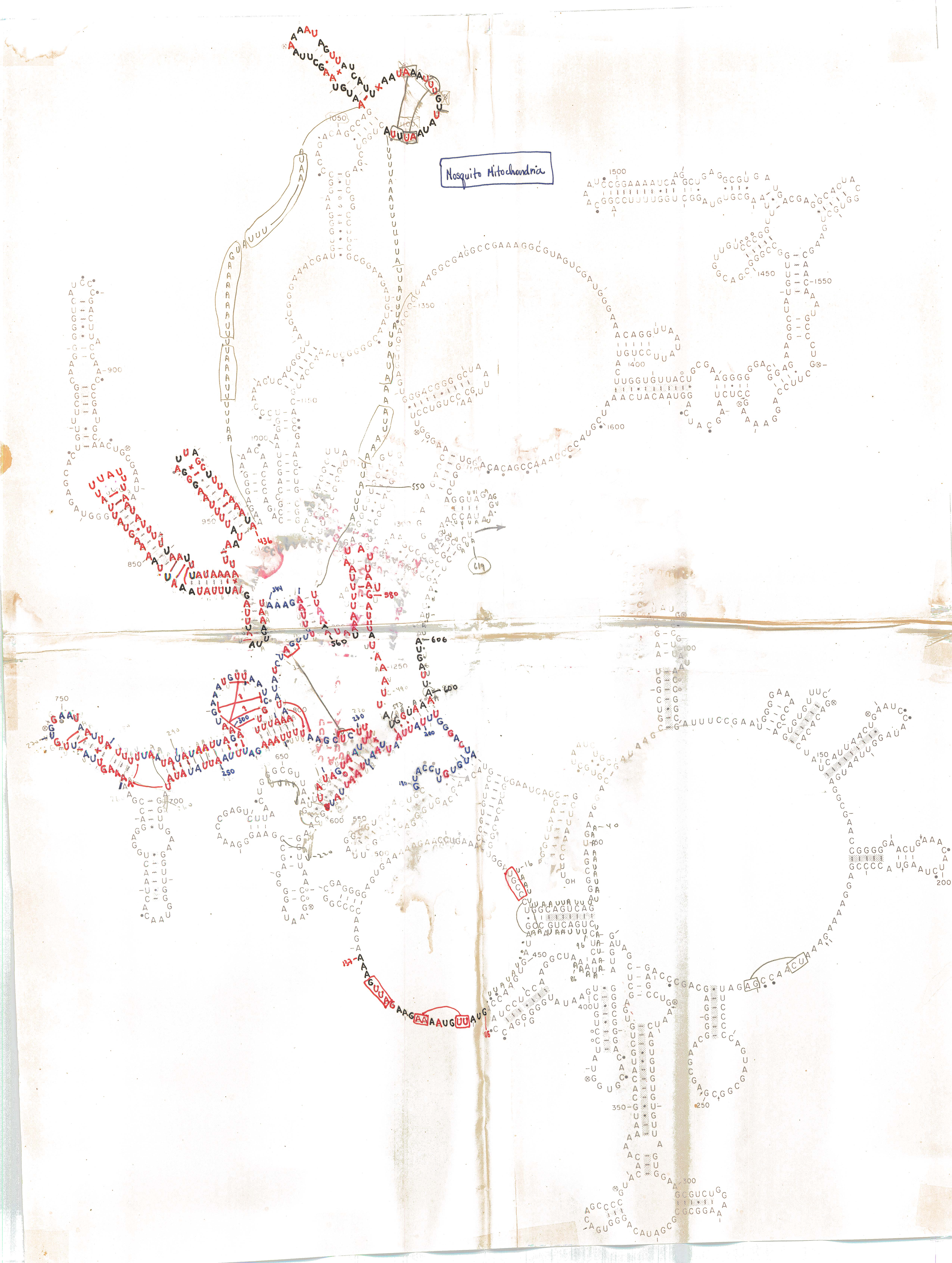
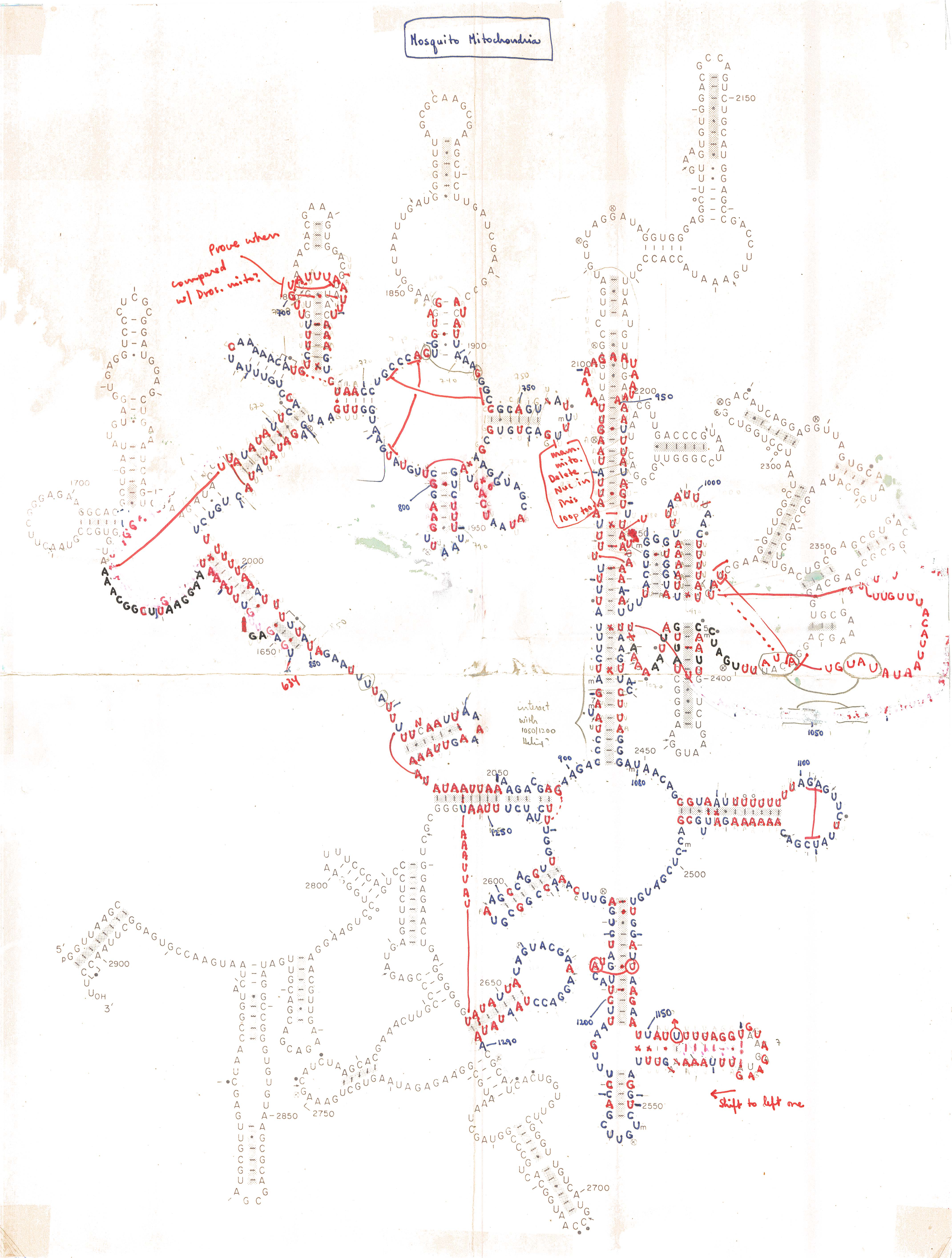
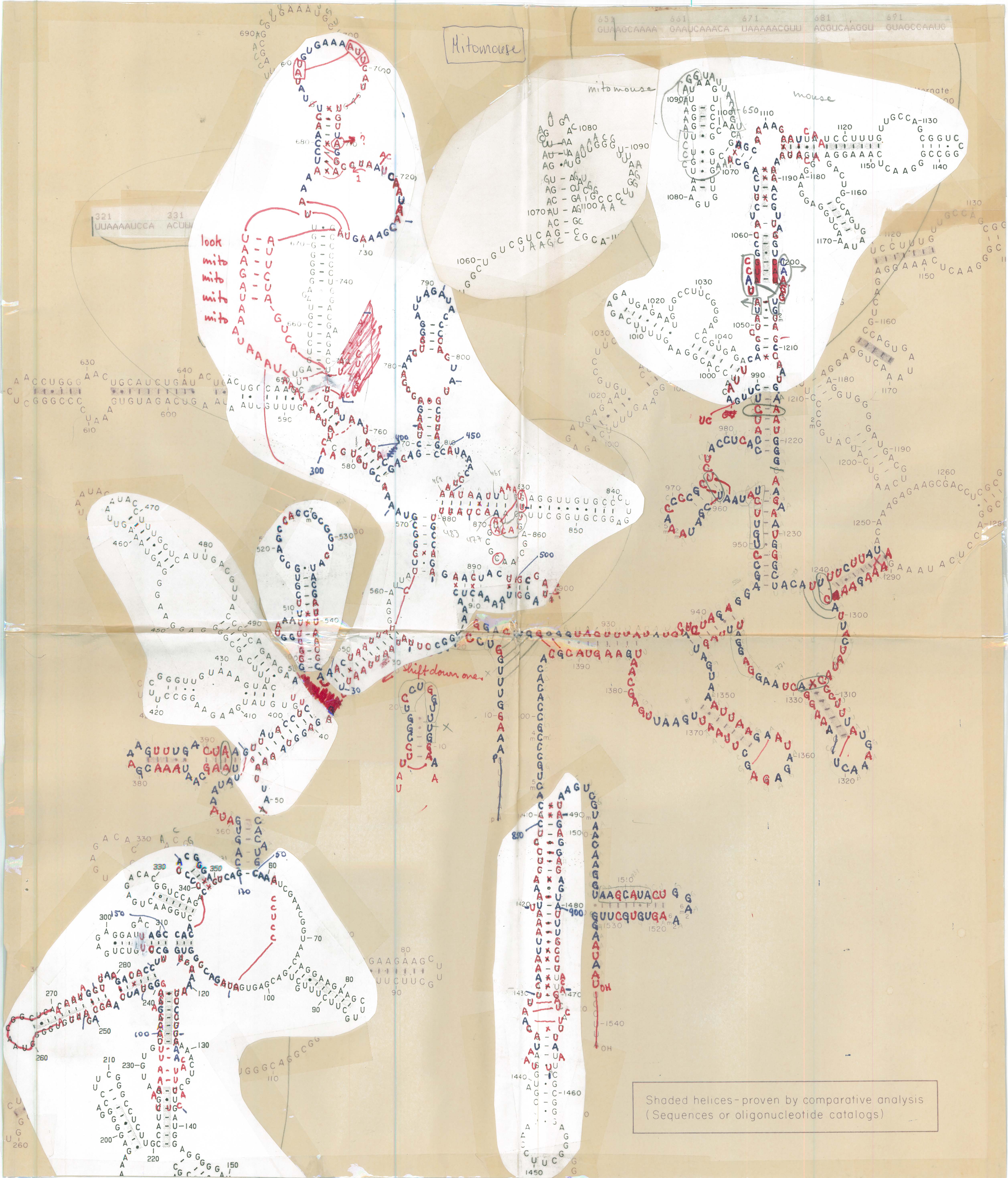
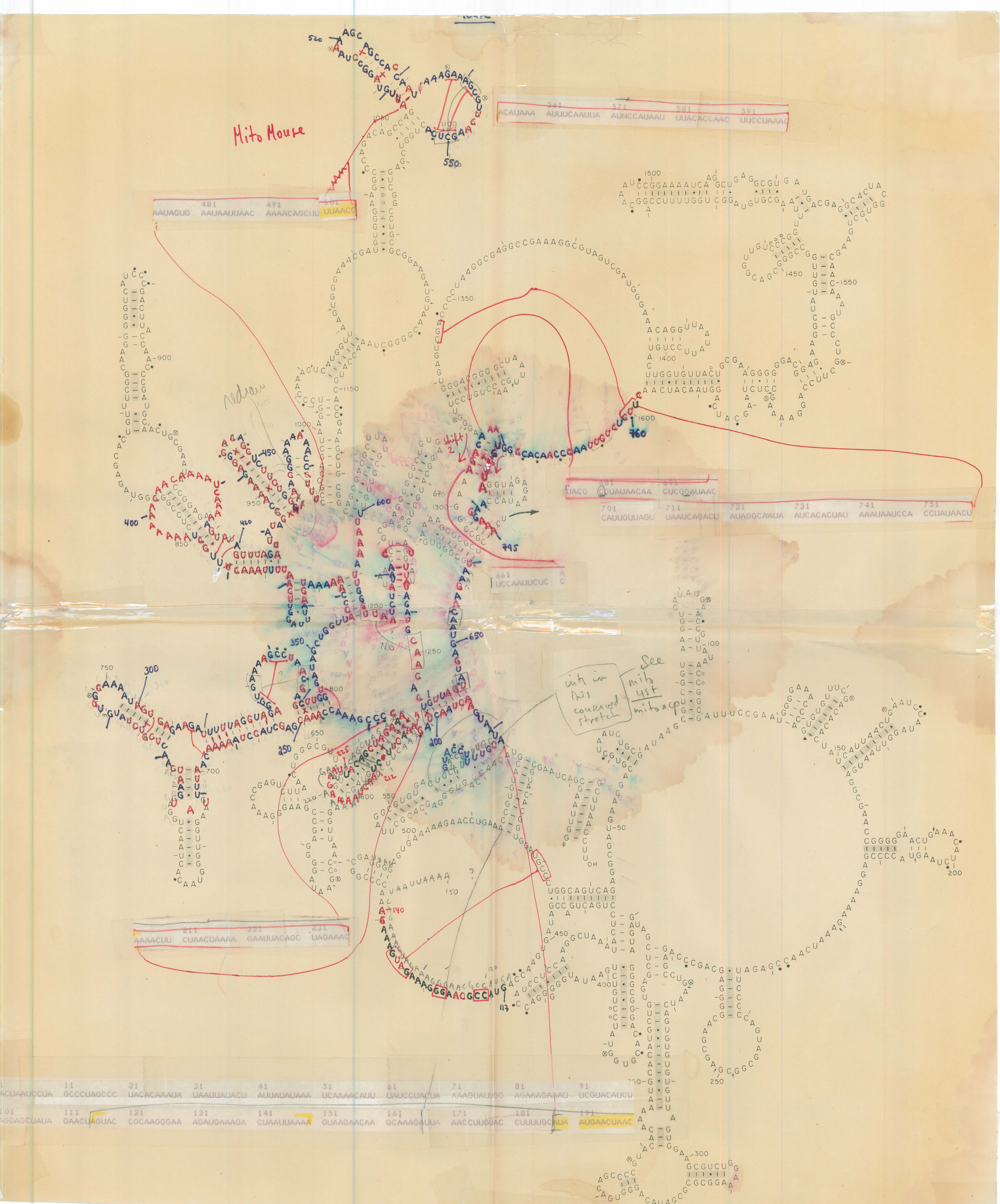
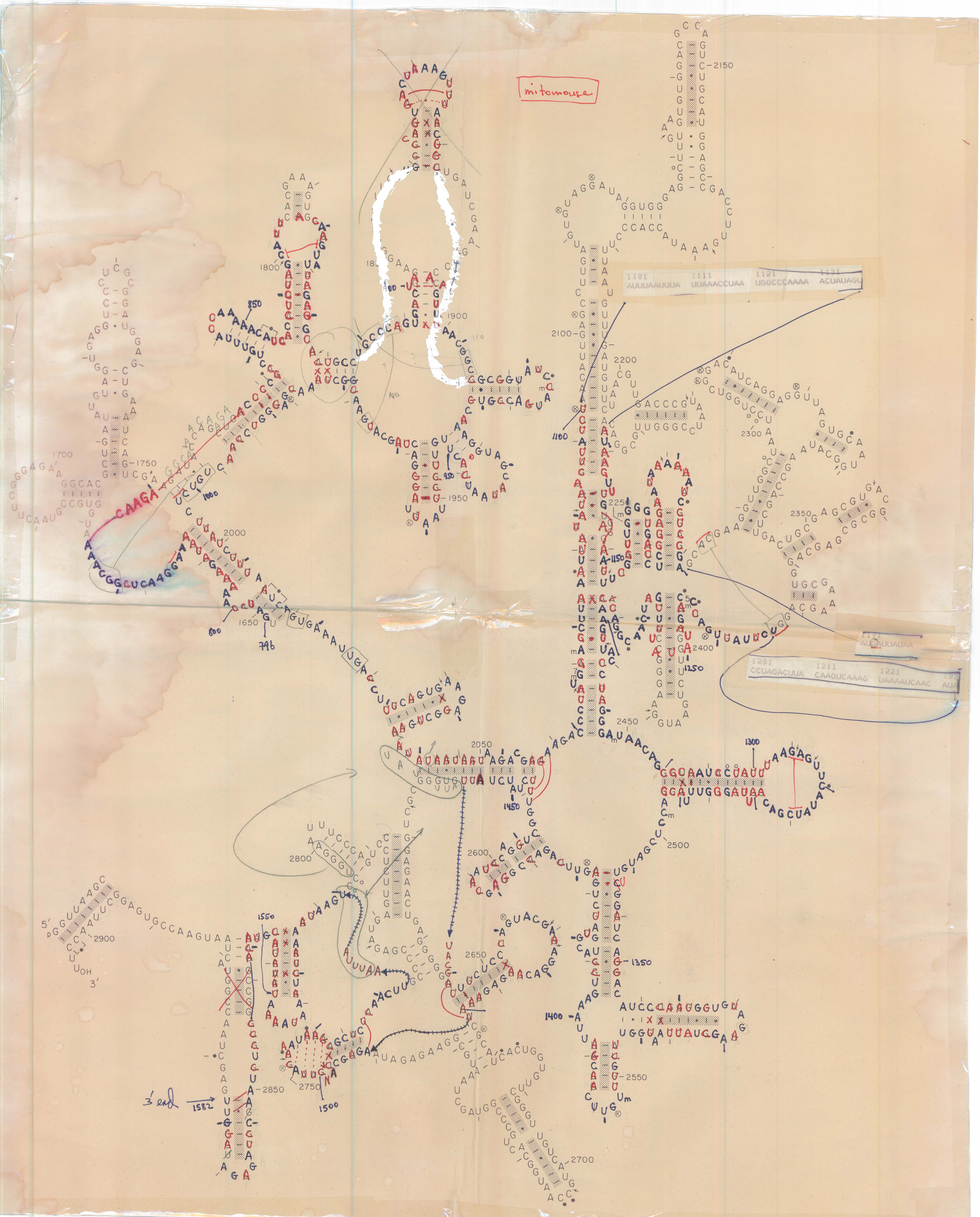
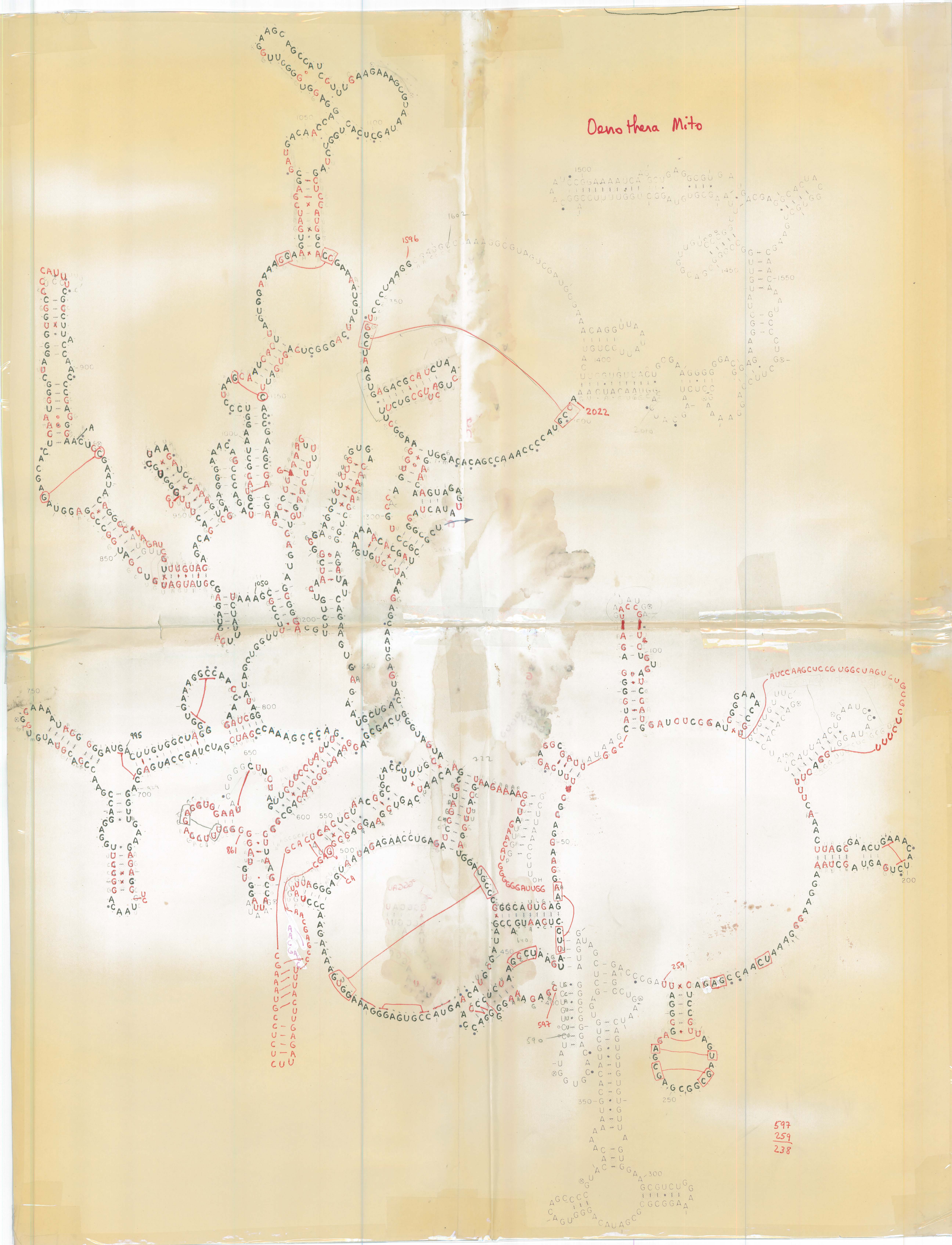
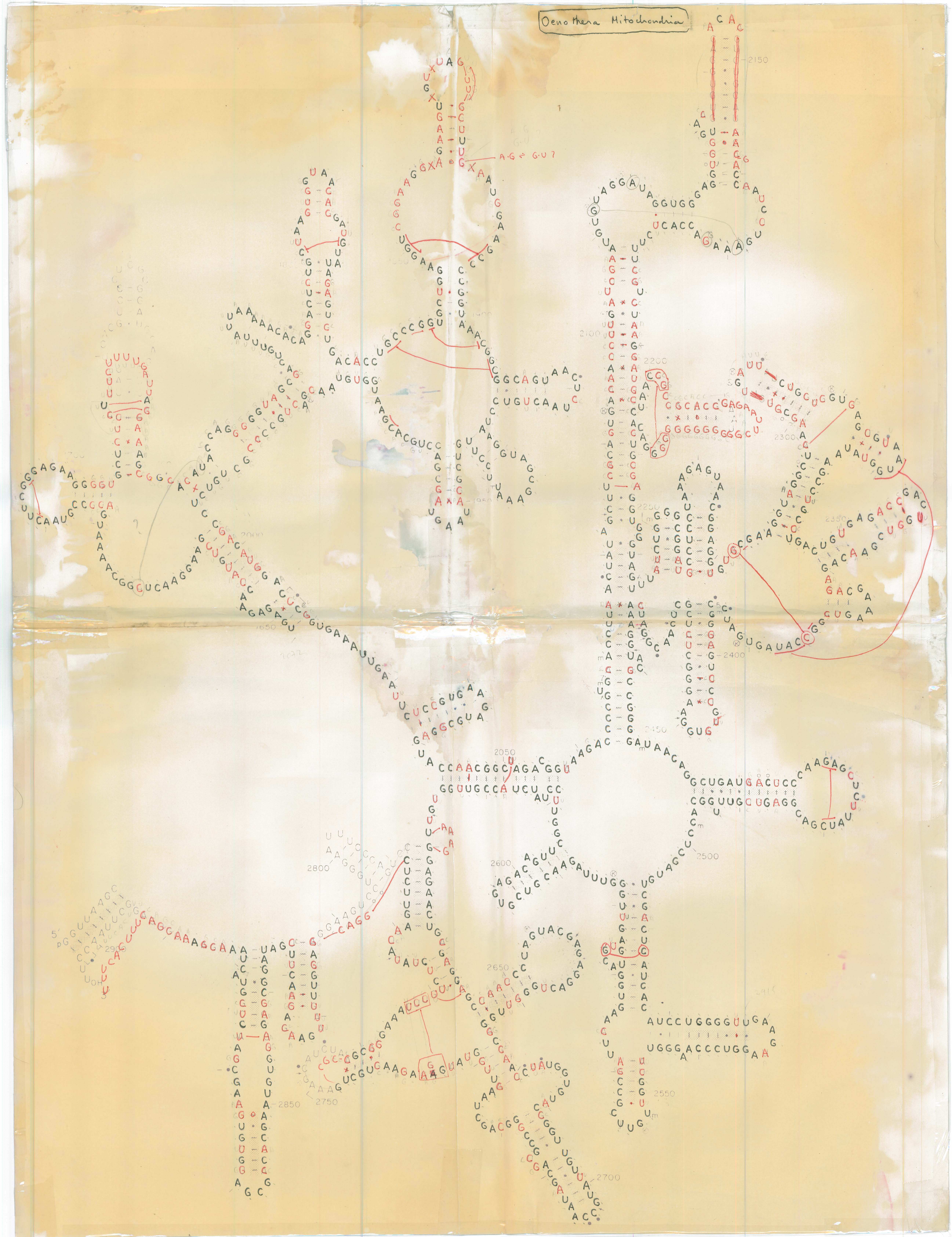
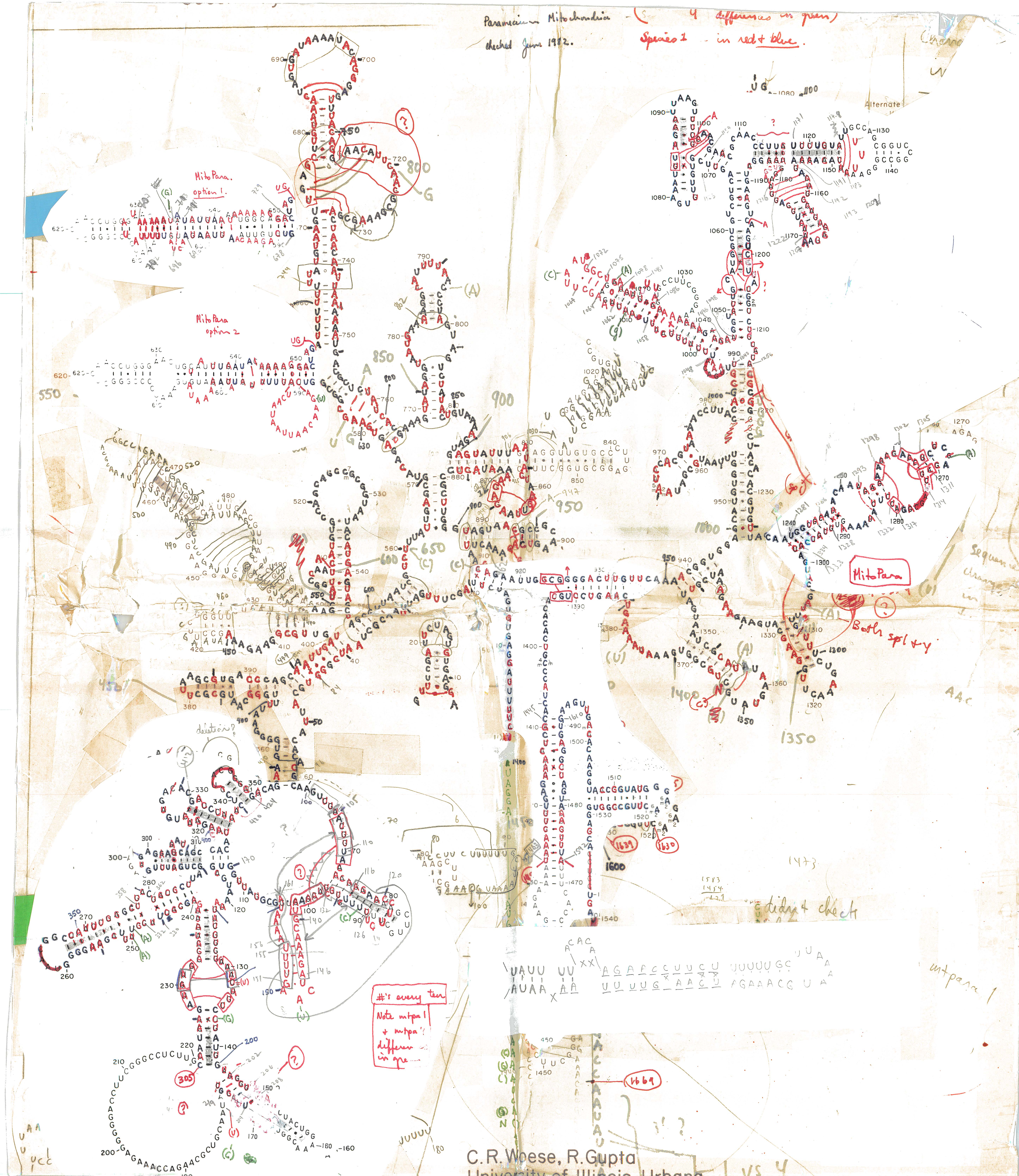
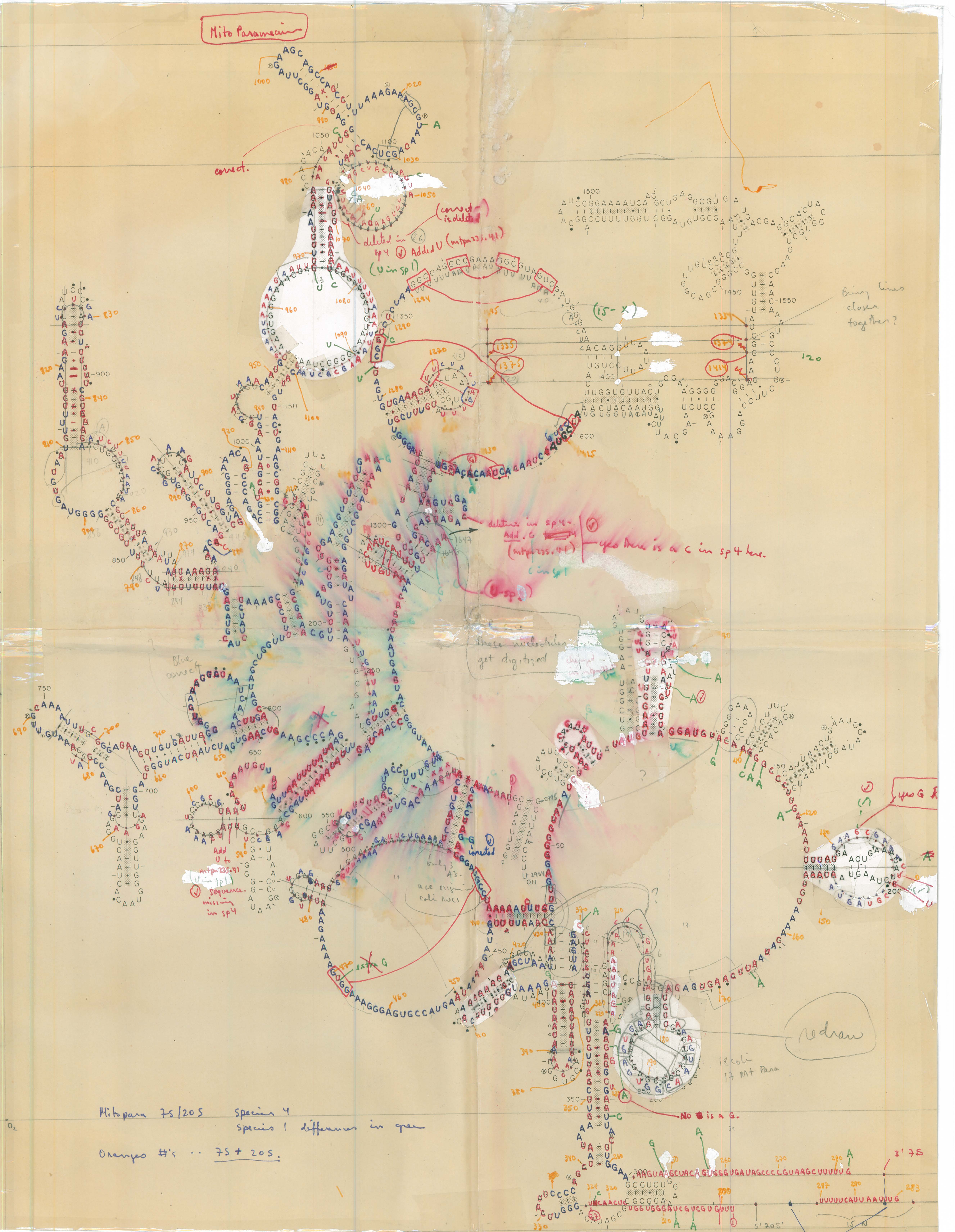
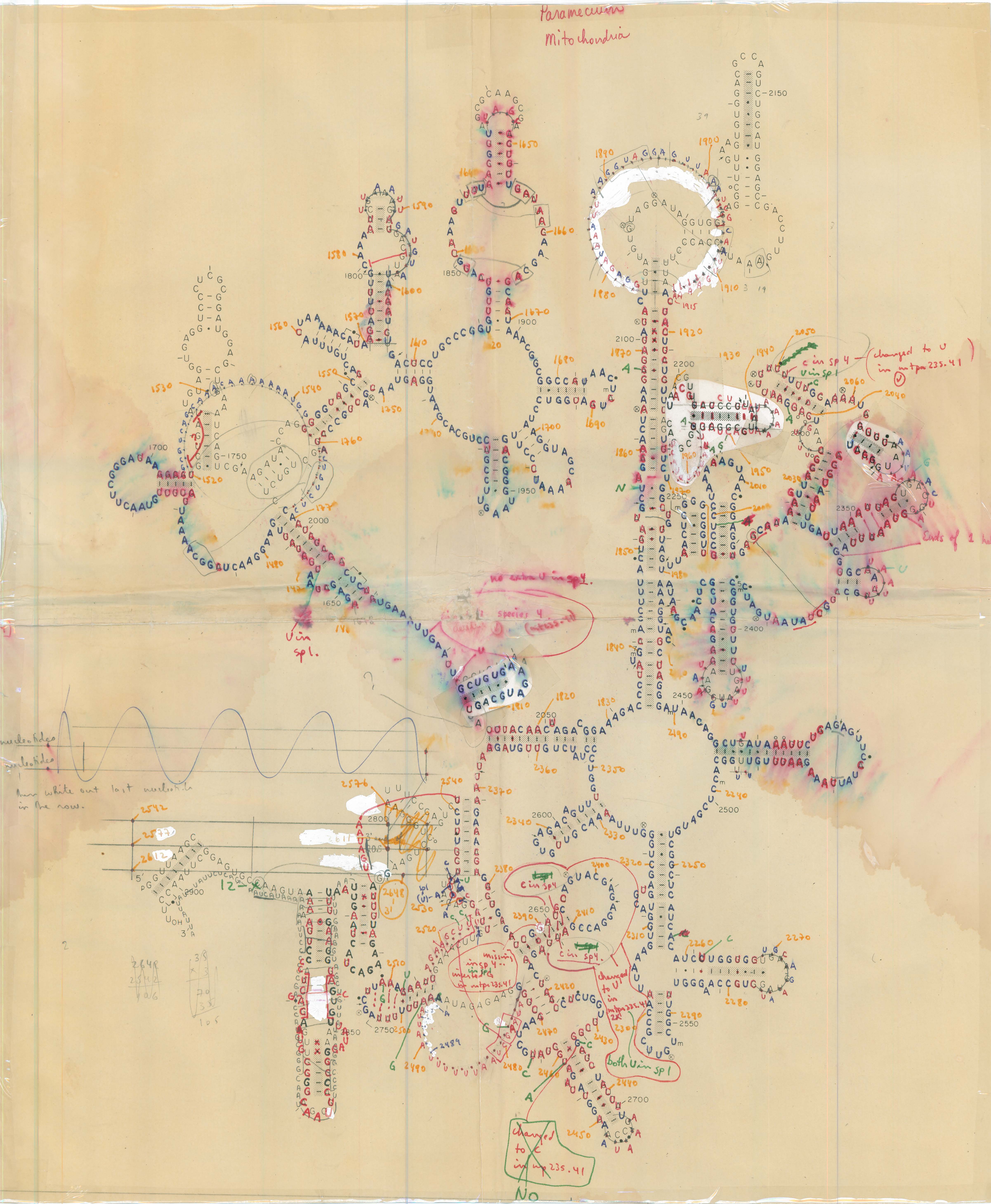
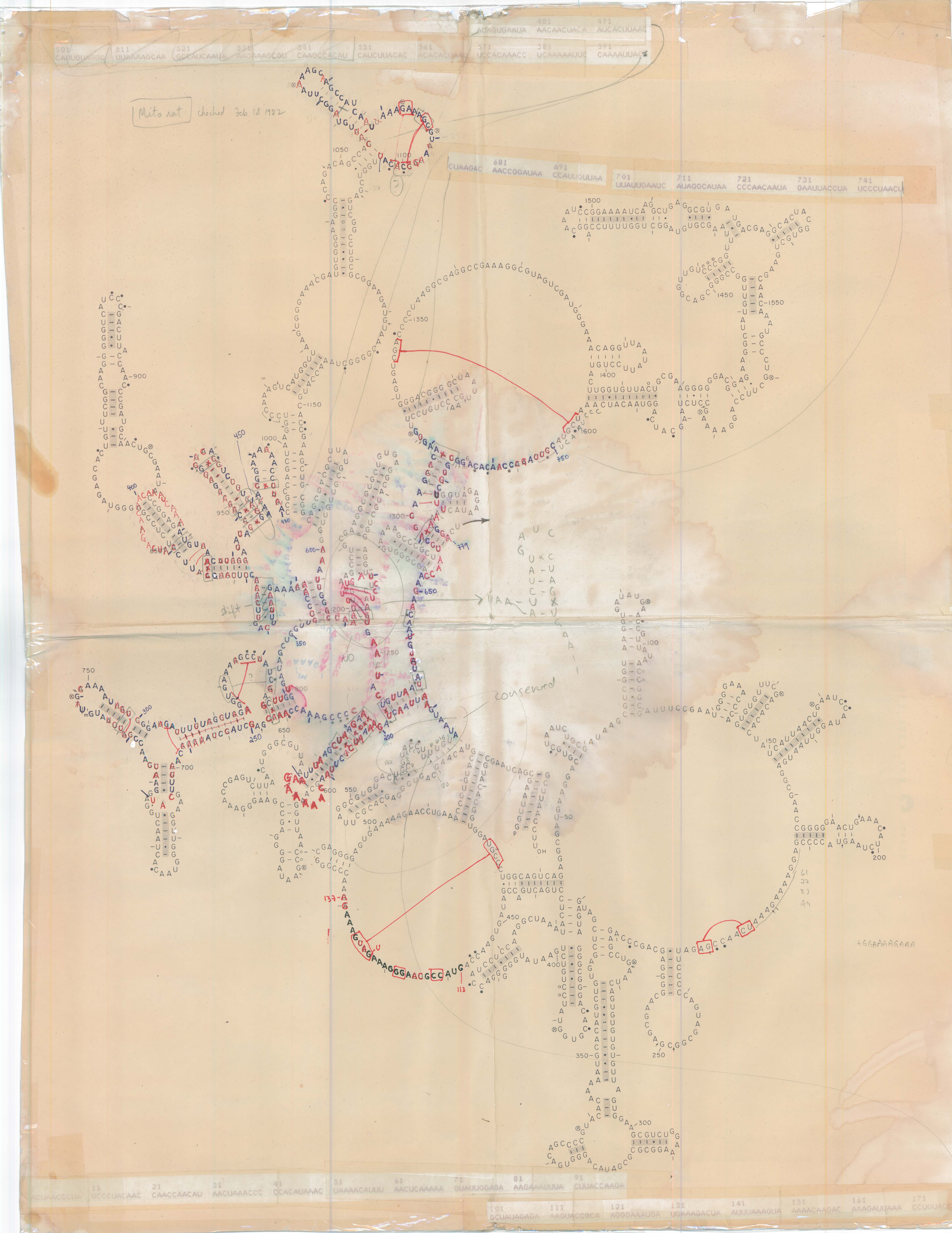
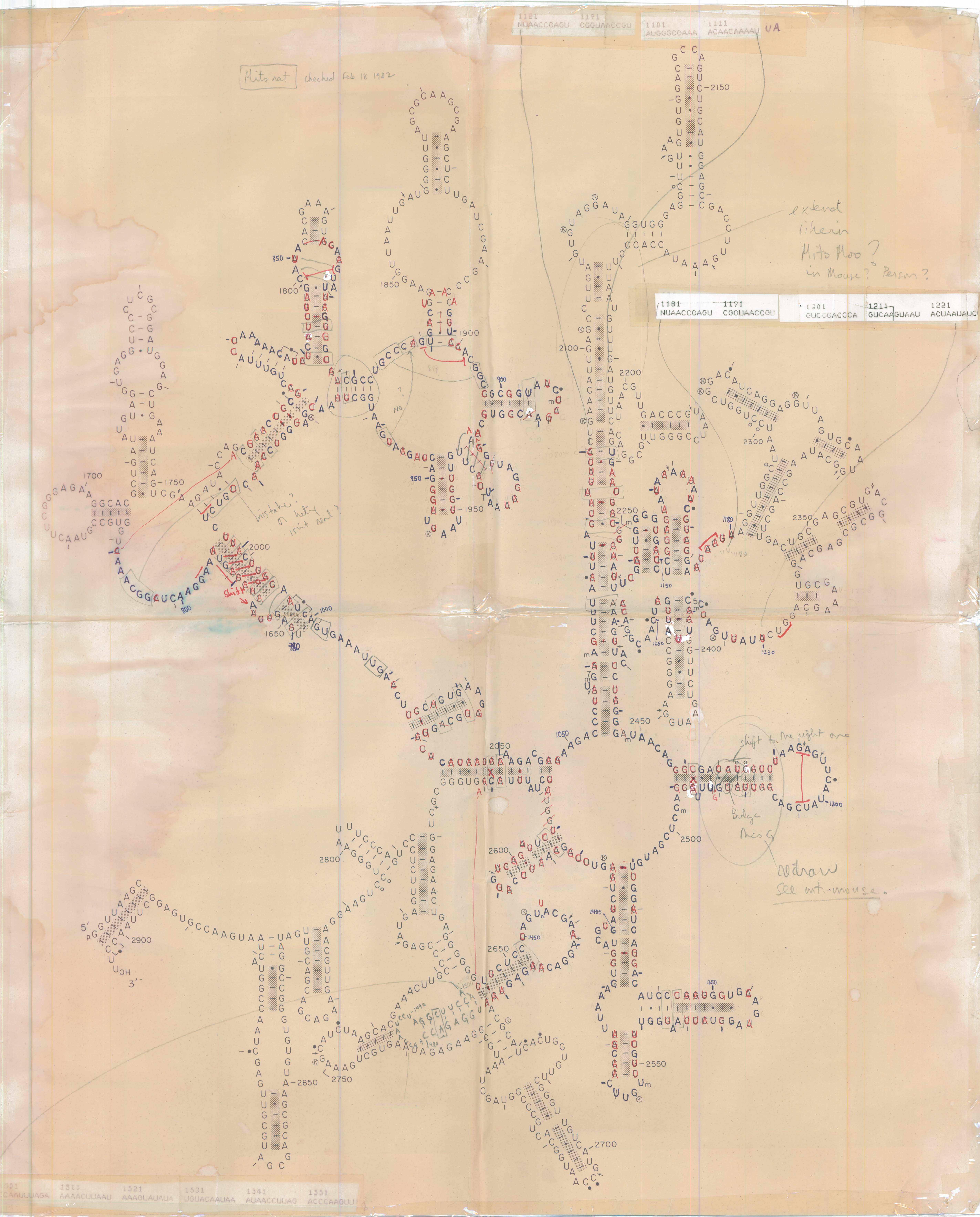
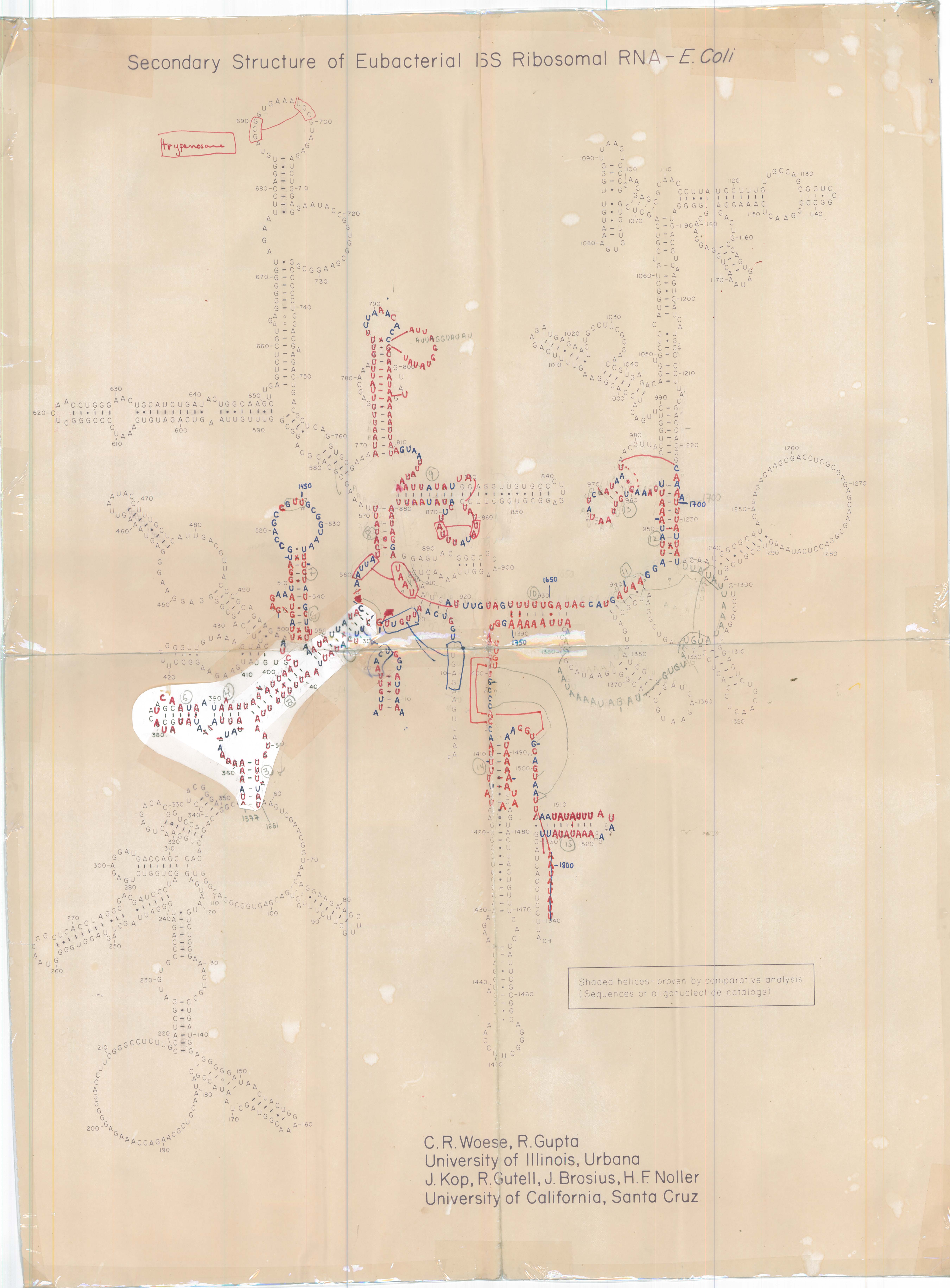
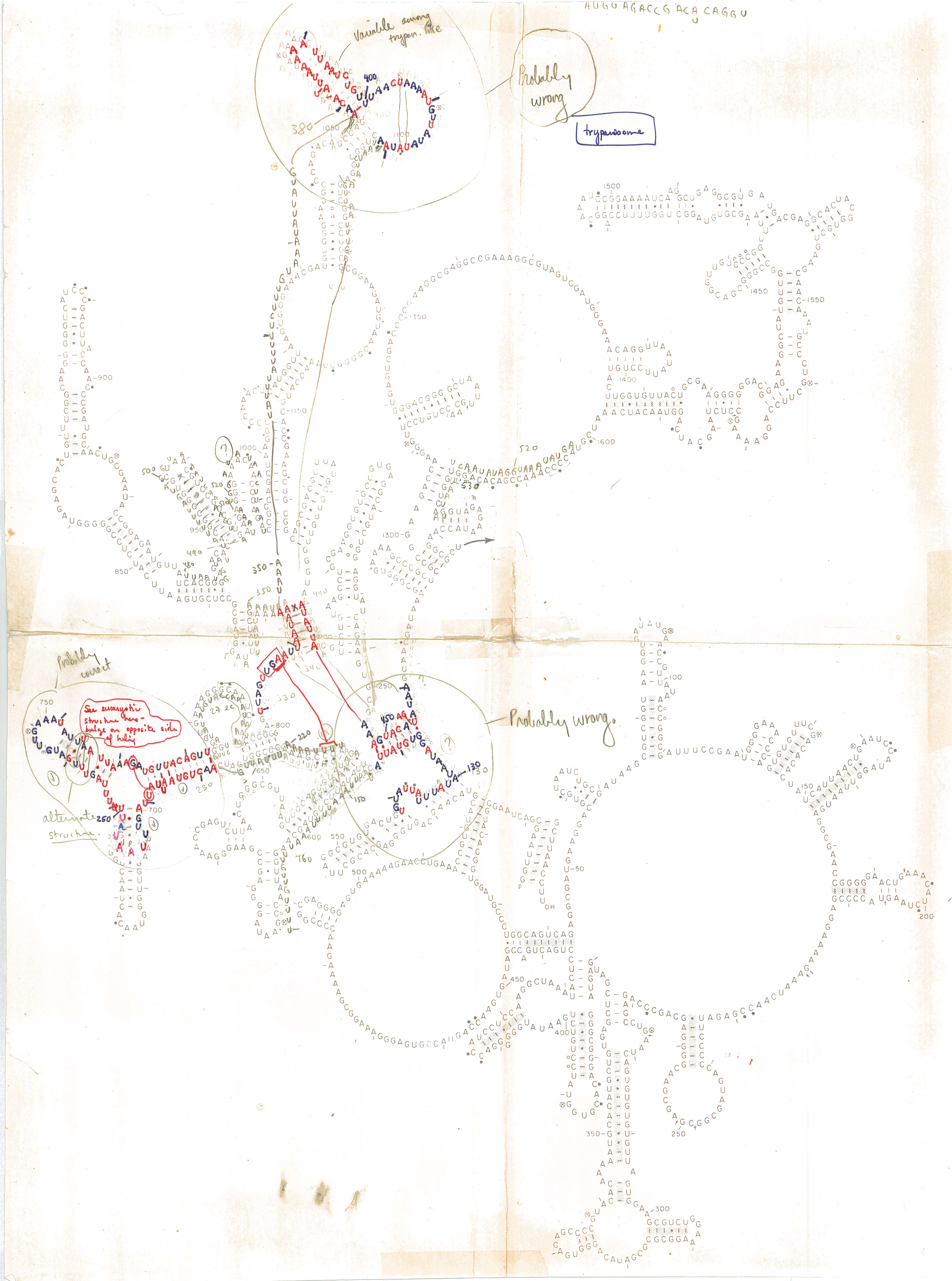
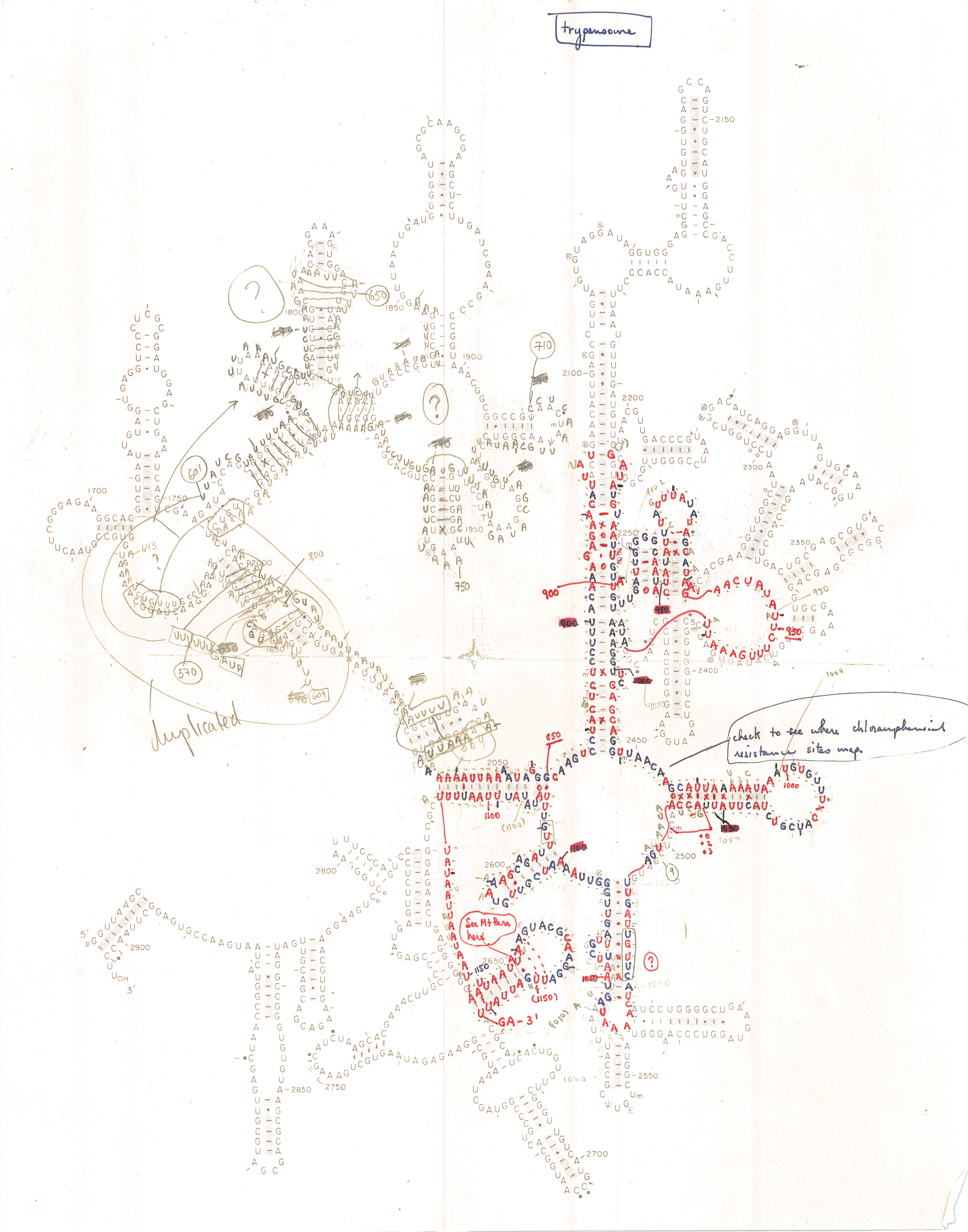
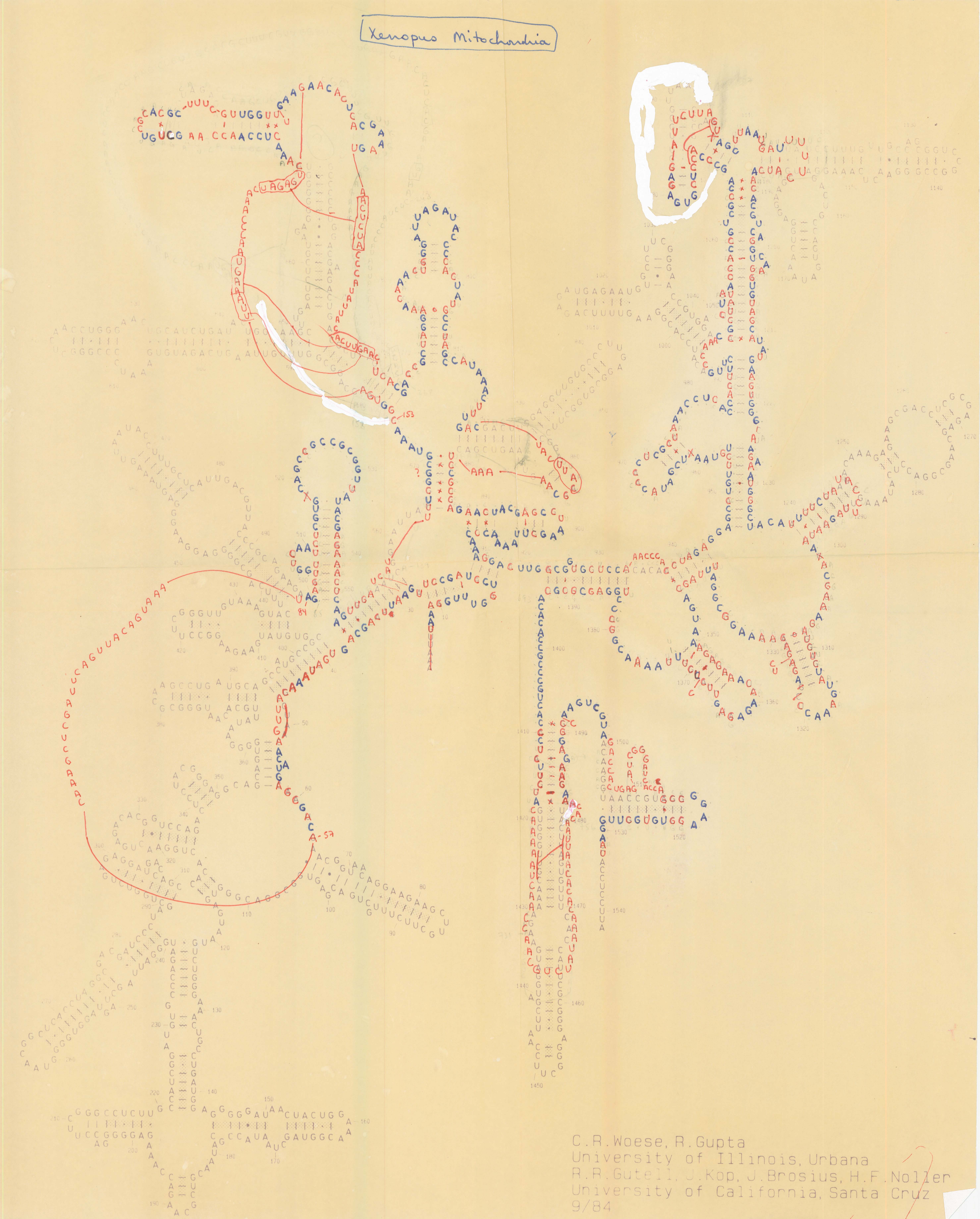
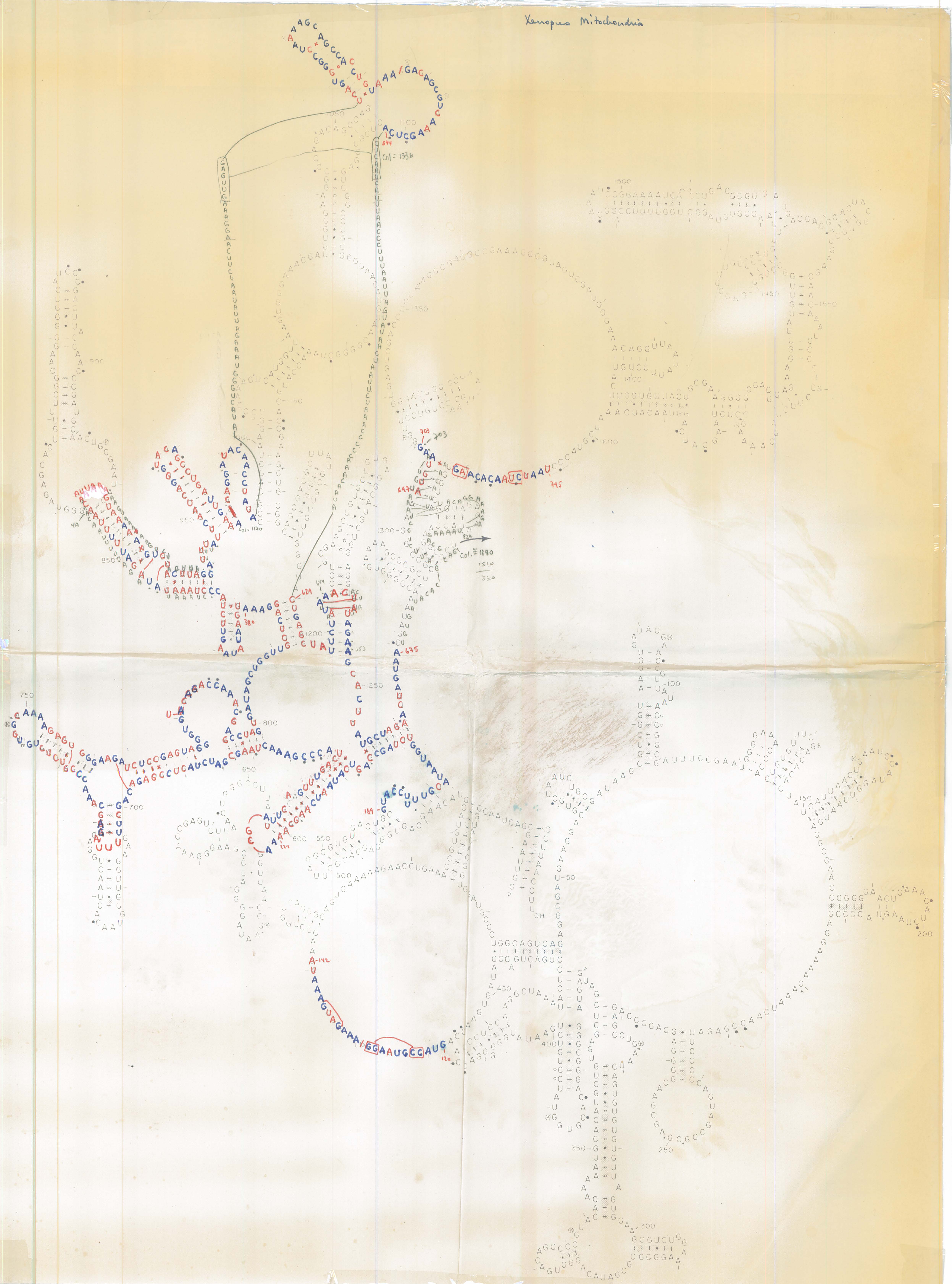
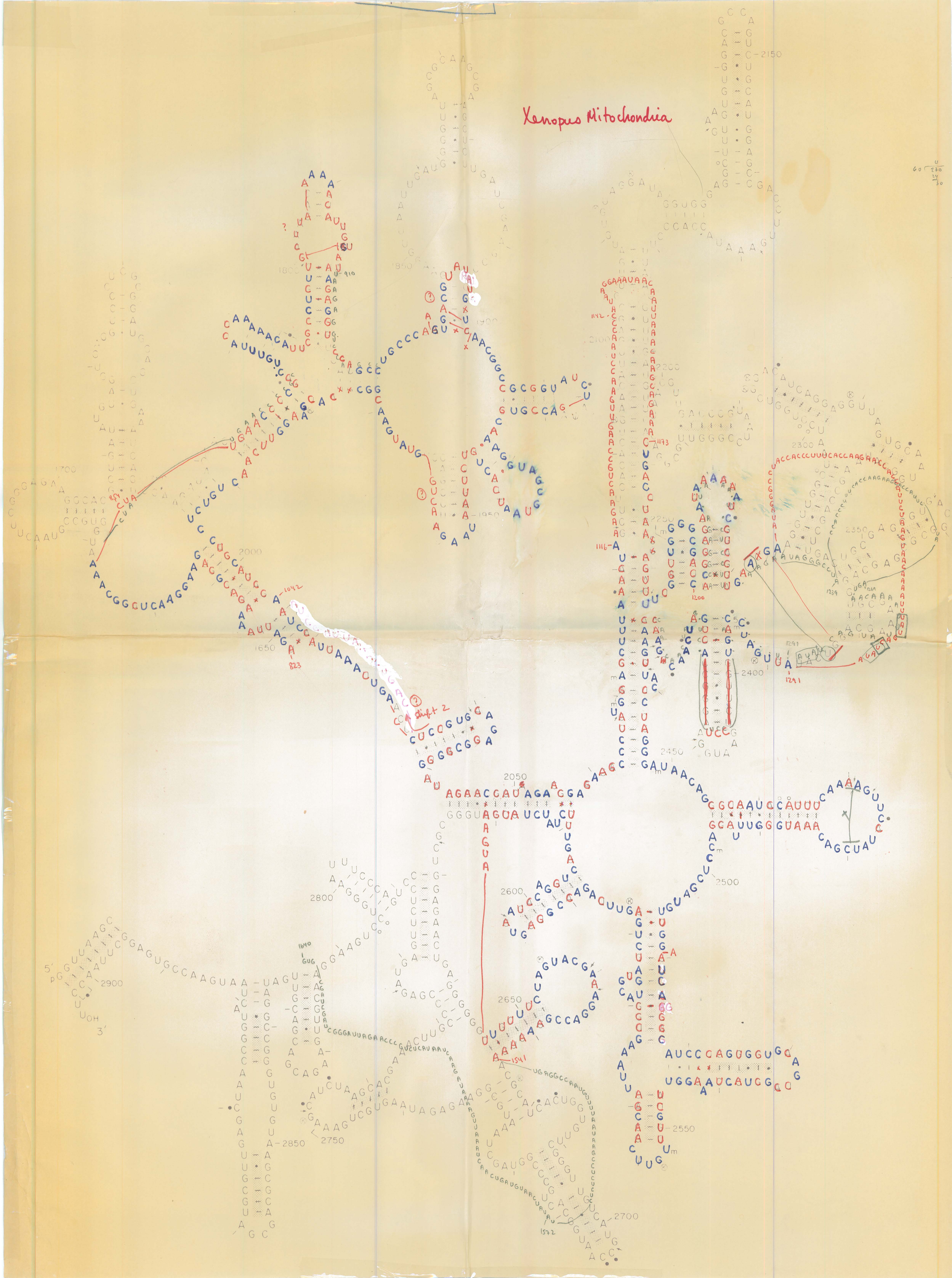
Carl Woese, in collaboration with Harry Noller, created large wall-size blueprints of the Escherichia coli 16S and 23S rRNAs secondary structure diagrams. Sequences from the 16S (and 16S-like) and 23S (and 23S-like) from other organisms were drawn, mapped, and threaded onto smaller versions of these diagrams. Initially, the second sequence was drawn over the E. coli sequence in pencil (see Bacterial MSB8 23S rRNAs), then finalized with blue letters to indicate similarity in nucleotides between E. coli and the other rRNA sequence, and red letters to indicate differences. Note that most base pairs in a helix are composed of two blue nucleotides or two red nucleotides (covariation). These hand drawings on blueprints were subsequently drawn with computers [4, 5, 10].
References:
Links to PubMed (PM), PubMed Central (PMC), and DOI are provided, when available.
- Woese C.R., Magrum L.J., Gupta R., Siegel R.B., Stahl D.A., Kop J., Crawford N., Brosius J., Gutell R., Hogan J.J., and Noller H.F. (1980). Secondary structure model for bacterial 16S ribosomal RNA: phylogenetic, enzymatic and chemical evidence. Nucleic Acids Research, 8(10):2275-2293. [ PM | PMC | DOI ]
- Noller H.F., Kop J., Wheaton V., Brosius J., Gutell R.R., Kopylov A.M., Dohme F., Herr W., Stahl D.A., Gupta R., and Woese C.R. (1981). Secondary structure model for 23S ribosomal RNA. Nucleic Acids Research, 9(22):6167-6189. [ PM | PMC | DOI ]
- Woese C.R., Gutell R., Gupta R., and Noller H.F. (1983). Detailed analysis of the higher-order structure of 16S-like ribosomal ribonucleic acids. Microbiological Reviews, 47(4):621-669. [ PM | PMC | doi ]
- Gutell R.R. (1984). Comparative Structural Analysis of 16S Ribosomal-RNA. Dissertation: University of California, Santa Cruz; 1984.
- Gutell R.R., Weiser B., Woese C.R., and Noller H.F. (1985). Comparative anatomy of 16S-like ribosomal RNA. Progress in Nucleic Acid Research and Molecular Biology, 32:155-216. [ PM | pmc | DOI ]
- Woese C.R., Winker S. and Gutell R.R. (1990). Architecture of Ribosomal RNA: Constraints on the sequence of Tetra-loops. Proceedings of the National Academy of Sciences (USA), 87(21):8467-8471. [ PM | PMC | DOI ]
- Woese C.R. and Gutell R.R. (1989). Evidence for several higher order structural elements in ribosomal RNA. Proceedings of the National Academy of Sciences (USA), 86(9):3119-3122. [ PM | PMC | DOI ]
- Gutell R.R. and Woese C.R. (1990). Higher order structural elements in ribosomal RNAs: Pseudo-knots and the use of non-canonical pairs. Proceedings of the National Academy of Sciences (USA), 87(2):663-667. [ PM | PMC | DOI ]
- Gutell R.R., Noller H.F., and Woese C.R. (1986). Higher order structure in ribosomal RNA. The EMBO Journal, 5(5):1111-1113. [ PM | PMC | doi ]
- Gutell R.R. and Fox G.E. (1988). A compilation of large subunit RNA sequences presented in a structural format. Nucleic Acids Research, 16(Supplement):r175-r269. [ PM | PMC | doi ]
rRNA Secondary Structure Model Diagrams:
NOTE: Some of these image files are large enough that heavily-loaded web browsers may not display them. You may wish to view this page in a new browser window and open links in new browser windows to avoid this problem.
| Taxonomy/Cell Location | Organism | SSU rRNA | LSU rRNA, 5' half | LSU rRNA, 3' half |
|---|---|---|---|---|
| Archaea | Desulfurococcus mobilis | JPG | ||
| Archaea | Halobacterium cutirubrum | JPG | ||
| Archaea | Halococcus morrhua | JPG | ||
| Archaea | Haloferax volcanii | JPG | JPG | JPG |
| Archaea | Methanobacterium formicium | JPG | ||
| Archaea | Methanococcus vannielli | JPG | JPG | JPG |
| Archaea | Methanospirullum | JPG | ||
| Archaea | Pyrodictium | JPG | ||
| Archaea | Sulfolobus solfataricus | JPG | JPG | JPG |
| Archaea | Thermococcus celer | JPG | ||
| Archaea | Thermoplasma acidophilum | JPG | ||
| Archaea | Thermoproteus tenax | JPG | ||
| Bacteria | Anacystis nidulans | JPG | JPG | |
| Bacteria | Agribacterium | JPG | JPG | |
| Bacteria | Bacillus stearothermophilus | JPG | JPG | |
| Bacteria | Bacillus subtilis | JPG | JPG | |
| Bacteria | Escherichia coli | JPG | JPG | |
| Bacteria | MSB8 | JPG | JPG | |
| Eukaryotic (Nuclear) | C. elegans | JPG | JPG | |
| Eukaryotic (Nuclear) | Dictyostelium discoideum | JPG | JPG | JPG |
| Eukaryotic (Nuclear) | Maize | JPG | ||
| Eukaryotic (Nuclear) | Mouse | JPG | JPG | |
| Eukaryotic (Nuclear) | Physarum | JPG | JPG | |
| Eukaryotic (Nuclear) | Rat liver | JPG | JPG | JPG |
| Eukaryotic (Nuclear) | Rice | JPG | JPG | |
| Eukaryotic (Nuclear) | Saccharomyces cerevisiae (Yeast) | JPG | JPG | JPG |
| JPG | JPG | |||
| Eukaryotic (Nuclear) | Tetrahymena boreali | JPG | ||
| Eukaryotic (Nuclear) | Tetrahymena thermophila | JPG | ||
| Eukaryotic (Nuclear) | Variamorpha necatrix | JPG | ||
| Eukaryotic (Nuclear) | Xenopus | JPG | JPG | JPG |
| Eukaryotic (Chloroplast) | Chloroplast Maize | JPG | JPG | |
| Eukaryotic (Chloroplast) | Chloroplast Tabacco | JPG | JPG | |
| Eukaryotic (Mitochondrion) | Aspergillus | JPG | JPG | JPG |
| Eukaryotic (Mitochondrion) | Cow | JPG | JPG | JPG |
| JPG | ||||
| Eukaryotic (Mitochondrion) | Crithidia | JPG | ||
| Eukaryotic (Mitochondrion) | Drosophila | JPG | JPG | |
| Eukaryotic (Mitochondrion) | Homo sapiens | JPG | JPG | JPG |
| Eukaryotic (Mitochondrion) | Maize | JPG | JPG | |
| Eukaryotic (Mitochondrion) | Mosquito | JPG | JPG | |
| Eukaryotic (Mitochondrion) | Mouse | JPG | JPG | JPG |
| Eukaryotic (Mitochondrion) | Oenothera | JPG | JPG | |
| Eukaryotic (Mitochondrion) | Paramecium | JPG | JPG | JPG |
| Eukaryotic (Mitochondrion) | Podospora | JPG | JPG | JPG |
| Eukaryotic (Mitochondrion) | Rat | JPG | JPG | JPG |
| Eukaryotic (Mitochondrion) | Trypanosome | JPG | JPG | JPG |
| Eukaryotic (Mitochondrion) | Xenopus | JPG | JPG | JPG |
| Eukaryotic (Mitochondrion) | Yeast | JPG | JPG | JPG |
Intron Secondary Structure Model Diagram:
| Intron RNA Molecule | Taxonomy/Cell Location | Organism | Intron RNA |
|---|---|---|---|
| Group I Intron | Eukaryotic Nuclear | Tetrahymena thermophila | JPG |