
Last modified on 16 August 2002.
Literature Reference:
Mears J.A., Cannone J.J., Stagg S.M., Gutell R.R., Agrawal, R.K., and Harvey S.C. (2002)
Modeling a Minimal Ribosome Based on Comparative Sequence Analysis.
Journal of Molecular Biology, 321:215-234.
Manuscript Figures and Tables:
| Figure/Table Legend | Panel |
|---|---|
| Table 1. Comparative analysis. | ( HTML ) |
| Table 2. Protein conservation. | ( HTML ) |
| Table 3. Deletions in the C. elegans alignment. | ( HTML ) |
| Table 4. Bridges conserved through evolution. | ( HTML ) |
| Table 5. Conserved tRNA/rRNA contacts within the ribosome. | ( HTML ) |
| Table 6. 16S and 23S rRNA Sequences Used for Comparative Analysis. | ( HTML ) |
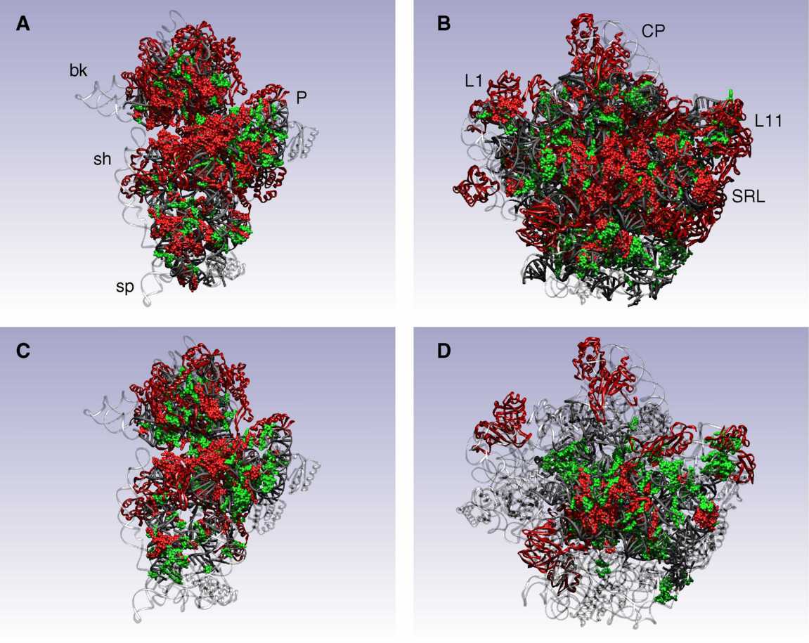
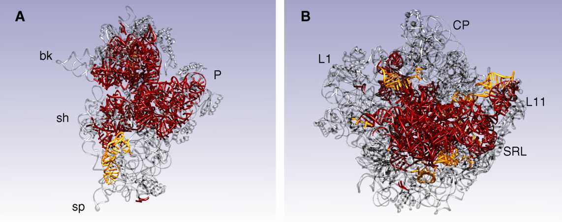
| Figure 1. Conserved regions of the ribosome. A, 3P / SSU; B, 3P / LSU; C, 3P2O / SSU; D, 3P2O / LSU. | ( JPG ) |
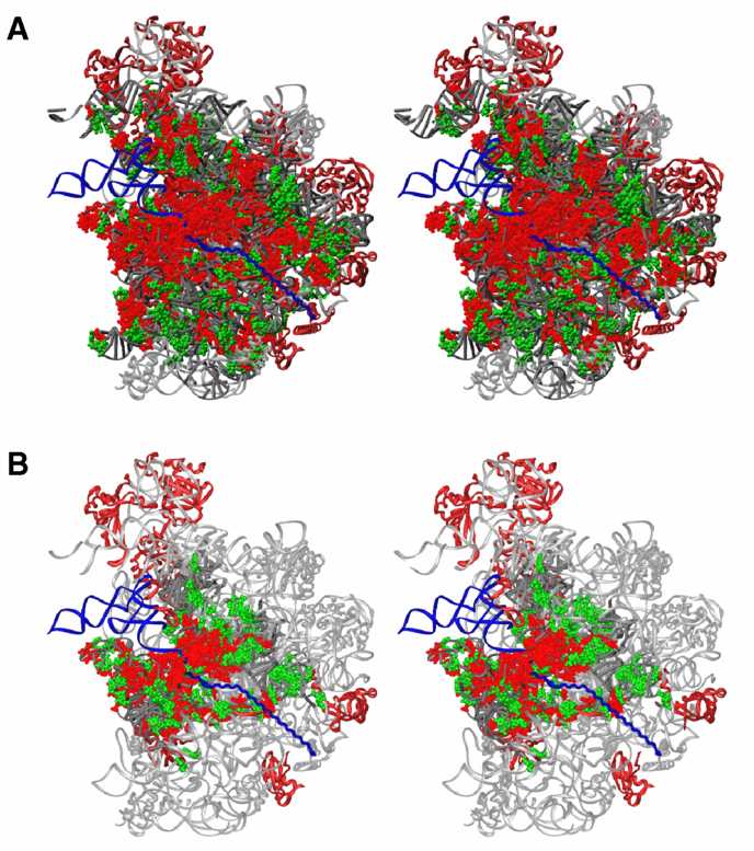
| Figure 2. The polypeptide-conducting tunnel. A, 3P; B, 3P2O. | ( JPG ) |
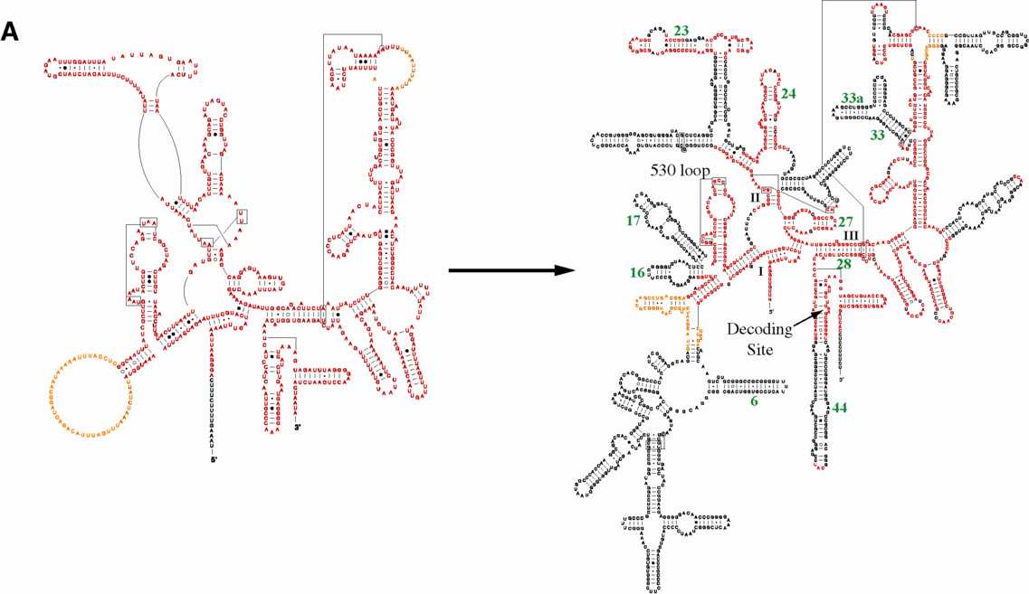
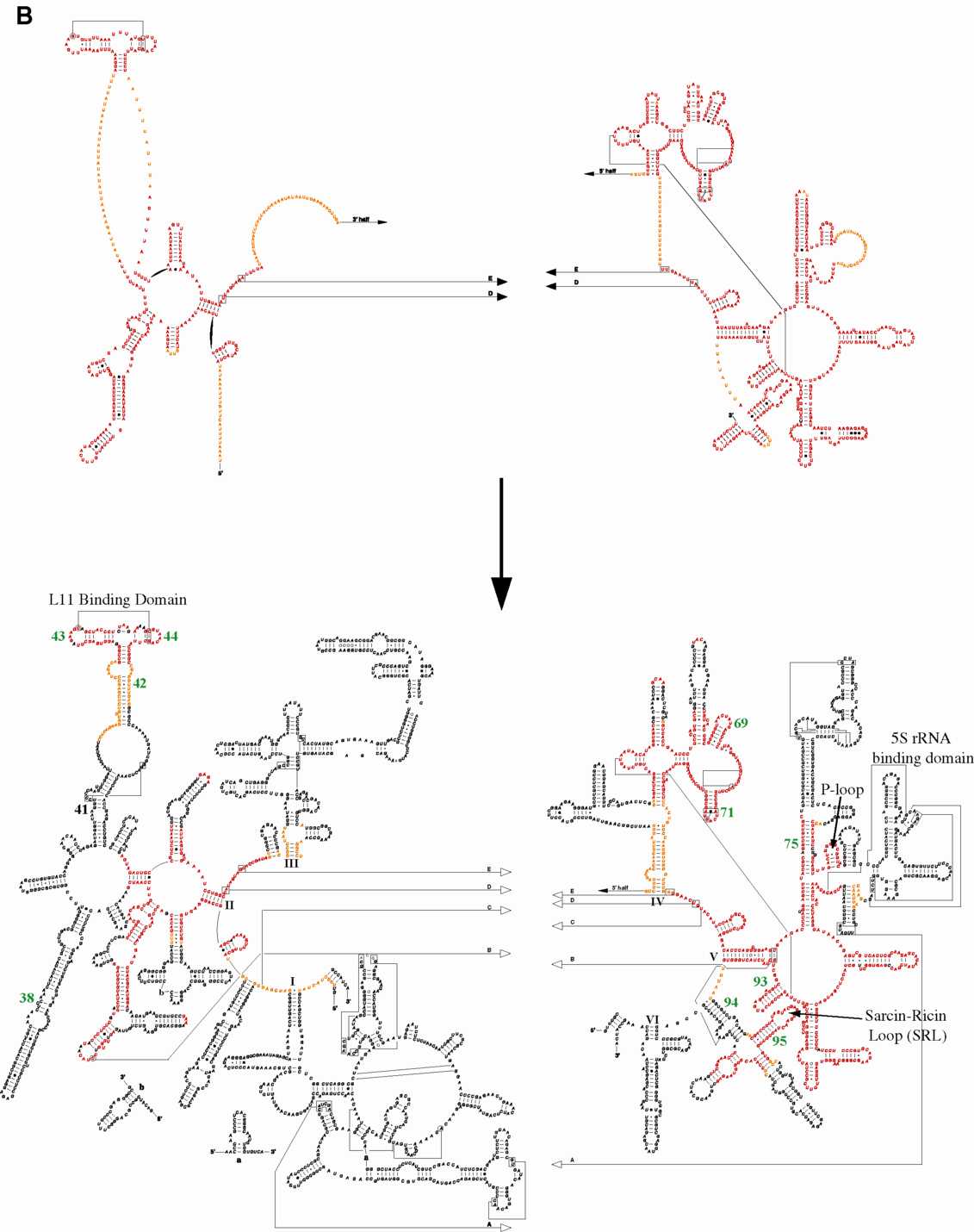
| Figure 3. C. elegans alignment mapped onto secondary structures. | A. SSU ( JPG | PS ) B. LSU ( JPG | PS ) |
| Figure 4. C. elegans alignment mapped onto crystal structures. A, SSU; B, LSU. | ( JPG ) |
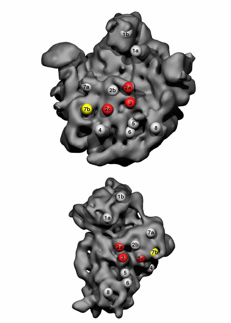
| Figure 5. Positioning of bridge contacts. | ( JPG ) |
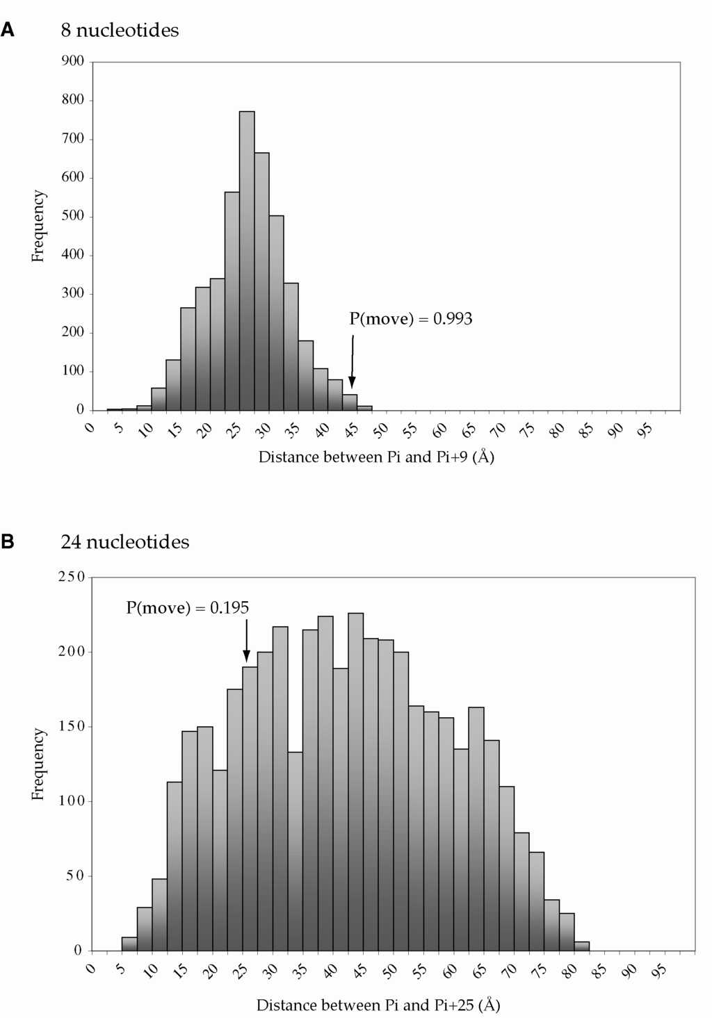
| Figure 6. Probability of movement. A, LSU Helix 94; B, LSU Helices 47-61. | ( JPG ) |
Additional Figures and Tables:
| tRNA/mRNA Binding Site vs. rRNA Conservation | ( List of Positions ) | ( Data Table ) |
| rRNA Nucleotide Numbering/Conservation Tables: | 16S rRNA: ( CONS.5 | CONS.1 ) | 23S rRNA: ( CONS.5 | CONS.1 ) |
rRNA Conservation Diagrams:
| Phylogenetic Group | 16S rRNA | 23S rRNA |
|---|---|---|
| Three Phylogenetic Domains | ( PS | PDF ) | 5' half: ( PS | PDF ) -- 3' half: ( PS | PDF ) |
| Three Phylogenetic Domains/Two Organelles | ( PS | PDF ) | 5' half: ( PS | PDF ) -- 3' half: ( PS | PDF ) |