
Highlights / Data
Contains the most recent comparative secondary structure model diagrams for each molecule's reference sequence, as well as alternative formats for the standard model diagrams.
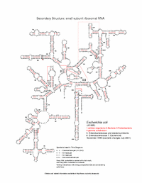
- Nucleotide: basic secondary structure model diagram, showing the strength of the comparative support information for each interaction.
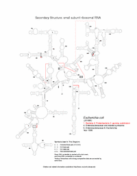
- Tentative Model: contains interactions with some comparative support, but not enough for inclusion in the final models.
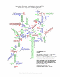
- Helix Numbering: basic secondary structure diagram showing the names of the helices that correspond with the helix names in the nucleotide frequency tables.
- Schematic: basic secondary structure model diagram, with the nucleotides replaced with a line traversing the RNA backbone.
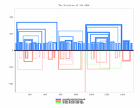
- Histogram: the RNA sequence is displayed as a single line from left (5') to right (3'), with three line segments connecting the two positions that form the secondary and tertiary structure base pairs.
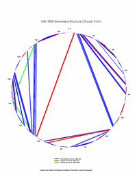
- Circular: the RNA sequence is drawn clockwise (5' to 3') in a circle, starting at the top. A single straight line connects the positions that form secondary and tertiary structure base pairs.
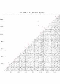
- All Possible Helices: All possible helices composed of four or more consecutive G:C, A:U and/or G:U base pairs are drawn on an X/Y plot.
| Reference | Helix Numbering | Schematic | Histogram | Circular | All Possible |
|---|---|---|---|---|---|
 |
 |
 |
 |
 |
 |